By Ashley M. Bancroft, DMD, Henderson, Nev; and James J. Bancroft, DMD, Waldwick, NJ
Postorthodontic white spots are the result of tooth structure’s exposure to cariogenic bacteria that reside in the plaque accumulation on the surfaces of the teeth and around brackets. An innovative therapy for cariogenic white spots has been developed that allows for the treatment of these unsightly lesions in a single office visit. The technique is known as resin infiltration, but has become synonymous with the product used for the treatment: Icon from DMG America. We have been using the Icon product successfully in our office over the past year.
What Causes the White Spots?
As enamel breaks down during the demineralization process, a subsurface network of channels begins to form within the enamel. As the lesion progresses, these networks grow deeper and more complex, then fill with an air/fluid mixture.
White-spot lesions are caused by the difference in the refraction of light as it passes through the air-fluid mixture of the lesion, as contrasted with the normal refraction through healthy enamel. The lesion’s refractive index bends light differently from the index of healthy enamel, and so appears whitish in color.
Clinical Treatment Steps
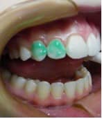
A 15-year-old female presented with postorthodontic decalcification lesions on the facial surfaces of #6, 7, 10, and 11. None of the lesions were cavitated. (Figure 1).

We cleaned the surfaces of each tooth with pumice, then placed a liquid dam (Opal Dam by Ultradent) to protect the gingiva from the etchant (Figure 2).
It is necessary to remove a thin layer of remineralized enamel that covers most white-spot lesions. This removal provides access for the infiltrating resin to flow freely into the lesion. If the treatment area is etched with a 15% HCl etchant (Icon Etch) for 2 minutes, this removes the surface layer of the lesion body, exposing the porosites of the lesion. We then rinse the tooth for 30 seconds and repeat the etching process. This second etching step is key to the successful outcome of the case.
After the treatment site has been thoroughly dried with oil-free and water-free air for 15 seconds, we apply a chemical drying agent (Icon Dry) to the surface and leave it undisturbed for 30 seconds, which completely desiccates the lesion, creating an environment for capillary action to take place. Following this 30-second period, we dry the site using oil-free air.
Within seconds of applying the Icon Dry, there should be a visible change in the appearance of the lesion. The lesion should fade, and the area should take on a more aesthetic appearance. Should the lesion remain whitish or opaque, it should be etched a third time.


Resin Application
We apply the resin infiltrant (Icon) in two steps. We apply the infiltrant into the treatment area in small increments to ensure that there is a reservoir of material to be drawn from the surface into the lesion. The material will continue to absorb into the dried lesion while additional material is exuded from the syringe.
After 3 minutes, we clean the area of excess material with cotton rolls. We clear proximal contacts with dental floss to prevent bonding the contacts of the adjacent teeth. We then light-cure the resin material for a total of 40 seconds per lesion and place a second application of resin onto the treatment site, following the same steps as before. We then finish the surface with a fine abrasive cup or disk.
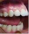
This procedure for resin infiltration of white-spot lesions has pleasing aesthetic results for the patient, parent, and orthodontist (Figure 3).


