Part 1 of a series designed to raise awareness of breathing problems and the procedures currently available to orthodontists to assist patients in improving their breathing, sleeping, and overall health.
By Robert “Tito” Norris, DDS
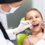
As members of the dental profession, orthodontists are in a unique position to recognize early signs and symptoms of upper airway obstruction and sleep disordered breathing (SDB). Three important elements of a new patient process can bring these indicators to light:
- Thorough health history including a specific questionnaire dedicated to breathing and sleeping;
- Comprehensive extraoral and intraoral clinical examination; and
- Radiographic examination.
Recognizing Upper Airway Obstruction and Sleep Disordered Breathing
The world is waking up to the importance of unobstructed breathing, particularly while sleeping. We have wearables, apps, beds, pods, pillows, drinks, medications, supplements, and literally hundreds of intraoral appliances designed to help us identify and improve our breathing while we sleep. Public awareness of the importance of SDB has never been higher. Therefore, patients and parents are quite amenable to engage in a conversation about this subject during a new patient examination (NPE). These conversations are an integral part of a holistic approach to orthodontic and dento-facial orthopedic care, and differentiate between those practitioners who desire to not only straighten teeth, but also improve the quality of life, health, and longevity of the patient. And this comprehensive approach begins with asking the right questions by having every new patient complete a sleep questionnaire.
Implementing Sleep Questionnaires in the Orthodontic Practice

Several standardized sleeping and breathing questionnaires have been developed by researchers in the specialties of sleep medicine and otorhinolaryngology (ENT). Unfortunately, there is not a global consensus as to which one is most comprehensive, effective, or diagnostic. Examples include the Epworth Sleepiness Scale (ESS),1 Pittsburgh Sleep Quality Index (PSQI),2 Sino-Nasal Outcome Test (SNOT-22),3 and Chervin Pediatric Sleep Questionnaire (CPSQ).4 After consulting with several sleep medicine specialists and ENTs in our area, our practice chose to implement the CPSQ (Figure 1) as a screening tool for all children and a SNOT-22 (Figure 2) questionnaire for all adults, as they are both validated patient-reported outcome measure (PROM) instruments.The SNOT-22 assesses 22 nasal, otologic, emotional, and sleep quality symptoms.

Since most new orthodontic patients are children, the CPSQ is the most commonly used screening tool in our practice. An increasing score on the CPSQ or SNOT-22 correlates with the severity of sleep disordered breathing. During the exam, we discuss any positive responses to those screening tools with the patient/parent and then review with them our clinical and radiographic findings in relation to those positive responses.
Correlating Questionnaire Scores with Clinical and Radiographic Findings

On the cephalometric radiograph, we first look at maxillo-mandibular positions in relation to the cranium (Figure 3). Retrusive maxillo-mandibular positions can be a key factor in SDB.5 The “ceph” also provides 2D visualization of the adenoid tissues and pharyngeal airway space (Figure 4). We then move on to our Cone Beam Computed Tomography (CBCT) findings, where we analyze the Minimal Axial Airway (MAA) (Figure 5). The probability of severe OSA is high with an MAA of less than 52 mm2, intermediate if the airway is 52 to 110 mm2, and low if the airway is more than 110 mm2, according to an article in the Journal of Oral and Maxillofacial Surgery.6 However, these values must be correlated to the symptoms revealed by the Sleep Questionnaire responses, as a CBCT is a static image taken in an erect position of dynamic tissues which are most often in a supine position while sleeping. From a transverse CBCT slice through the first molars (Figure 6), maxillary and mandibular alveolar widths can be compared.

This view also affords imaging of the nasal septum, turbinates, paranasal sinuses (PNS), and resting tongue position. A midline cleft or groove in the dorsal surface of the tongue can be indicative of a posterior tongue tie, and an air space between the tongue and the palate indicates low tongue posture. Low tongue posture can be the result of simply a habitual posture, an anterior or posterior tongue tie, or a constricted nasal airway resulting in habitual mouth breathing.
Comprehensive Clinical Examination for SDB Indicators
In addition to a typical orthodontic clinical examination, our clinical exam encompasses a number of observations to screen for SDB. These include a high or narrow palatal vault, anterior or posterior crossbites, worn dentition, abfraction lesions, and localized gingivitis due to anterior xerostomia. The clinical exam also includes assessment of the nares for adequate width and valvular collapse during inhalation, measurement of the upper lip lengths at the philtrum and commissures, measurement of upper lip dynamics, grading of the anterior tongue mobility using the Tongue Range of Motion Ratio (TRMR),7 examining for signs of a posterior tongue tie and/or tongue scalloping, grading of the palatine tonsils using the Brodsky grading scale of tonsil size,8 and grading of the Mallampati index.9
Discussing Findings and Making Referrals
All of these findings are then discussed with the patient/parent. More often than not, there is an element of a compromised airway which is contributing to the malocclusion, and our duty as health care providers is to educate our patients as to where these problems lie and make the appropriate referrals when indicated. In certain cases where Obstructive Sleep Apnea (OSA) or Upper Airway Resistance Syndrome (UARS) is suspected, a patient will be sent home with a Home Sleep Test (HST) for further screening. Results will be reviewed on the following day as they relate to the patient’s overall orthodontic treatment plan.
Constructing Effective Referrals to ENT and Sleep Physicians
Although as orthodontists, we are not qualified nor authorized to make a diagnosis of OSA or UARS, we can construct an influential referral to an ENT or sleep physician colleague who is educated to the notion that we, as physicians of the mouth, are in a unique position to identify early signs and symptoms of SDB. After all, the specific and most common structures involved with airway obstruction include those that we can evaluate:
- Nasal valve collapse
- Deviated septum
- Hypertrophic turbinates
- Nasal polyps
- Obstructed paranasal sinuses
- Narrow maxilla and associated nasal airway width
- Retrusive maxilla and/or mandible
- Hypertrophic pharyngeal tonsils (adenoids)
- Hypertrophic palatine tonsils
- Hypertrophic lingual tonsils
- Hypertrophic tongue due to excessive fat
- Tongue tie and subsequent muscular hypertrophy
Understanding the naso-pharyngeal anatomy and possible sources of obstruction is key in building a strong referral to our ENT and Sleep Medicine colleagues. They can then assist in further testing patients, diagnosing airway problems, and pharmacologically or surgically reducing or removing airway obstructions to reduce airway resistance.
These well-educated colleagues understand that SDB is a progressive disease, and that early intervention is in the best interest of the long-term health, cognitive development, athletic and academic performance, and longevity of the patient. OP
READ MORE: Check out Part 2 on Tranverse Treatment and Part 3 on Anterior-Posterior Solutions

Robert “Tito” Norris, DDS, is a board-certified orthodontist in private practice at Stone Oak Orthodontics in San Antonio, Texas. Norris, who lectures both nationally and internationally, holds several patents and trademarks, and is the inventor of the Norris 20/26 Passive Self-Ligating Bracket System.
References
- Johns MW. A new method for measuring daytime sleepiness: The Epworth Sleepiness Scale. Sleep 1991; 14(6):540-5.
- D J Buysse 1, C F Reynolds 3rd, T H Monk, S R Berman, D J Kupfer The Pittsburgh Sleep Quality Index: a new instrument for psychiatric practice and research. Psychiatry Res. 1989 May;28(2):193-213.
- Kennedy JL, Hubbard MA, Huyett P, et al.. Sino-nasal outcome test (SNOT-22): a predictor of postsurgical improvement in patients with chronic sinusitis. Ann Allergy Asthma Immunol 2013;111:246-251
- RD Chervin 1, K Hedger, JE Dillon, KJ Pituch Pediatric sleep questionnaire (PSQ): validity and reliability of scales for sleep-disordered breathing, snoring, sleepiness, and behavioral problems. Sleep Med. 2000 Feb 1;1(1):21-32
- R W Riley 1, N B Powell, C Guilleminault Maxillary, mandibular, and hyoid advancement for treatment of obstructive sleep apnea: a review of 40 patients J Oral Maxillofac Surg. 1990 Jan;48(1):20-6.
- Schendel, Stephen & Jacobson, Richard & Khalessi, Sadri. (2012). Airway Growth and Development: A Computerized 3-Dimensional Analysis. Journal of oral and maxillofacial surgery : official journal of the American Association of Oral and Maxillofacial Surgeons. 70. 2174-83.
- Audrey Yoon 1, Soroush Zaghi 2 3, Rachel Weitzman 4, Sandy Ha 5, Clarice S Law 6, Christian Guilleminault 7, Stanley Y C Liu 2. Toward a functional definition of ankyloglossia: validating current grading scales for lingual frenulum length and tongue mobility in 1052 subjects. Sleep Breath. 2017 Sep;21(3):767-775.
- Brodsky L Modern assessment of tonsils and adenoids. Pediatr Clin North Am 1989;36 (6) 1551- 1569
- Mallampati, SR; Gatt, SP; Gugino, LD; et al. (July 1985). “A clinical sign to predict difficult tracheal intubation: a prospective study”. Can Anaesth Soc J. 32: 429–34.


