A German study finds that magnetic resonance imaging (MRI) is a well-tolerated imaging modality for the diagnosis of dental abnormalities in children and for orthodontic treatment and surgical planning. Moreover, when compared with conventional radiography, dental MRI provides the advantage of three-dimensionality and complete elimination of ionizing, which is particularly relevant for repeated examinations in children.
The study appears in the July issue of the Journal of Oral and Maxillofacial Surgery.
Researchers from the University of Würzburg looked at 16 patients, with a mean age of 10.8 years, to assess the feasibility of MRI of dental abnormalities in children. Selected from 1,500 orthodontics patients, the selected patients included three with mesiodens, nine with supernumerary teeth other than mesiodens, one with germination, one with dilacerations, one with transmigration, and one with transposition. Three-dimensional (3D) images were taken on a 1.5-T MRI scanner using a 3D turbo spin echo sequence with voxel size of 0.8 x 0.8 x 1 mm. The measurement time was 4 to 5 minutes.
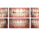
Using natural MRI contrast, the teeth, dental pulp, mandibular canal, and cortical bone could be clearly delineated. The position and shape of malformed teeth could be assessed in all three spatial dimensions.