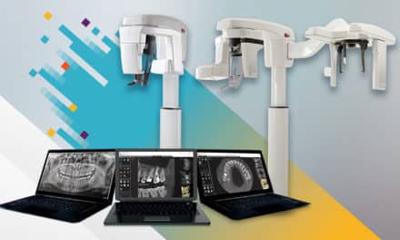
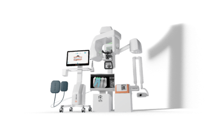
 X-rays are an established and necessary part of every orthodontic practice. I believe the quotes, "You don’t know what you don’t know," and, "You don’t know what you can’t see," pretty much say it all. CBCT offers views of the patient in all three dimensions with none of the magnification, distortion, or superimposition found in all traditional 2D x-rays. This precision allows diagnosis and treatment plans not obtainable with traditional 2D views. This diagnostic advantage is well demonstrated in the ability to evaluate the patient’s airway competence in a 3D model accurate to thousandths of an inch. Add to that the ability to surface-fuse an accurate 3D photogrammic image, and a complete hard- and soft-tissue analysis is available, including a three-dimensional analysis of the airway.
Consider the following radiation facts.
• Just being alive exposes a patient to .008mSv of background radiation every day.
• At an altitude of 30,000 feet, traveling from Los Angeles to London, a pilot or passenger is exposed to .080mSv. This exposure explains why the pilots were concerned with going through the scanners at airports. The exposure is also equal to 10 days of normal background radiation.
• A full-mouth series of small intraoral x-rays exposes the patient to .170mSv, or the equivalent of 22 days of background radiation.
• A medical CT of the skull (obtaining the same basic information as a CBCT scan) exposes the patient to .869mSv of radiation.
• If, for example, you have your gall bladder removed, a medical CT scan of the abdomen is standard at 10.000mSv of exposure—equivalent to 3 years of background radiation.
• A routine chest x-ray exposes the patient to .080mSv and, again, is the equivalent of 10 days of background radiation…
…As we start a discussion of 3D imaging, the first question that comes to mind is why image at all? One of the overall goals of orthodontic treatment should be to make the face more attractive.
—Joe Mayes, DDS, MSD
To read the rest of this article, click here.