by Sylvain Chamberland, DMD, MSc
The four steps to using this self-ligating bracket system
 |
| Sylvain Chamberland, DMD, MSc |
Self-ligating appliances were first introduced to the profession in the 1930s by Ford and Boyd.1 Over the next 40 years, many self-ligating designs were conceived but none resulted in any clinical or commercial impact. In the early 1970s, G. Herbert Hanson, DDS, began to work on the design of a new orthodontic bracket that would improve operator efficiency and achieve greater precision and control of tooth movement than previous designs had.
This led to the release of the SPEED bracket in the early ’80s.2 Since then, several modifications have made clinical use easier, but the fundamental design remains unchanged. One of the most significant modifications was the introduction of a superelastic nickel titanium spring clip.3 The clip maintains its stiffness and exerts consistent force on ligated archwires throughout treatment, while clips made of weaker material such as Elgiloy have shown extensive relaxation after use.4
Hanson’s design also offers the often-overlooked advantage of providing the user with a mix of so-called “passive” and “active” archwire interaction. Enhanced cooperation between the bracket and the archwire gives the user full 3D control over rotation, tip, and torque. Any deviation of the bracket position relative to the wire will result in deflection of the spring clip that will then seat the wire into its home position.
Bracket Placement
As with many other systems, precise bracket positioning is very important with the SPEED system. The basic placement principles are the same as with any straightwire edgewise appliance. The orthodontist determines the archwire plane by locating the maximum buccal convexity of the posterior teeth (Figure 1). This plane usually coincides with placing the slot of the bracket 4 mm to 4.5 mm from the incisal edges of the anterior teeth. A more incisal or gingival position of ± 0.5 mm may be decided according to the clinical crown length.
 |
| Figure 1: Selecting the archwire plane based on maximal buccal convexity. |
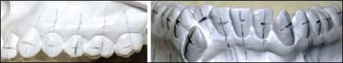
The user should consider two other parameters when placing the brackets: the long axis and the mesiodistal position of the bracket. The long axis of the anterior teeth is best assessed using the lingual and with a panoramic view (Figure 2). A meticulous bonding technique will pay dividends later in terms of efficiency and quality of treatment.
Archwire Progression
A typical archwire progression may be divided into four segments: initial alignment, arch levelling and torquing, tooth translation, and retraction and finishing. Each segment of treatment has different goals and thus requires different wire characteristics.
 |
| Figure 2: The long axis is assessed from the lingual for anterior teeth. Mesiodistal placement is on the middle lobe prominence of the teeth. |
- Initial alignment: The preferred wire at this stage of treatment is .016 or .018 SPEED Supercable™. Supercable contains multiple strands of superelastic nickel titanium wire. Regardless of malalignment, Supercable archwire provides 3 to 5 times less force than .014 or .016 solid nickel titanium wire.5
 |
| Figure 3: The initial archwire, a .016 Supercable, is left in place for 11 weeks. As unraveling occurs, the excess wire extends distally. |
The initial phase of treatment varies significantly from one patient (or malocclusion) to another, but generally it is accomplished in 6 to 18 weeks (Figure 3). The added value of taking time to align teeth in first and second order is that you might be able to skip a wire in the next phase.
- Arch leveling: The second phase of treatment consists of leveling the curve of Spee, coordination of the archform, and torquing. This should be done with rectangular NiTi archwire.
 |
| Figure 4: Note archform development from A at 21 weeks to C at 33 weeks. A .020 x .020 heat-activated NiTi wire is engaged 27 weeks into treatment (B) followed by a .020 x .025 SPEED nickel titanium wire 6 weeks later (C). |
The archwire sequence of this phase may include .016 x .022, .020 x .020, or .020 x .025 nickel titanium wire (Figure 4). Any significant bracket-placement errors that become apparent should be rectified at this stage before moving into heavier stainless steel wires.
This sequence should be followed by a .020 x .025 stainless steel SPEED archwire or .021 x .021 stainless steel D-wire. The goal would be to enhance torque, achieve archform coordination, and obtain a flat curve of Spee.
- Tooth translation and retraction: As mentioned above, en-masse retraction should be preceded by a full-dimensional, rigid archwire. This encourages full expression of the interaction between the superelastic spring clip, the archwire, and the archwire slot. The torque prescription will be expressed, and the curve of Spee will continue to flatten.
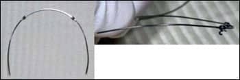 |
| Figure 5: A .021 x .021 x .018 Dual-geometry wire with reversed curve of Spee and posterior toe-in. If in the maxillary arch, the preactivation bend would accentuate the curve of Spee. |
En-masse retraction is achieved using a dual-geometry wire (Figure 5), square in the anterior (.021 x .021) and round in the posterior (.018 or .020). This configuration allows full torque control of the incisors during retraction, with significantly reduced friction in the posterior since the rounded part of the wire will be in the passive zone of the spring clip/archwire configuration.
When inserted into the arch, E-Links force module (TP Ortho) can be attached to the first or second molars according to the anchorage requirement. The force used to close the extraction space is applied above the center of resistance of the posterior unit and anterior unit in the sagittal view. The previously formed reversed curve of Spee creates a moment that will counteract the tipping of the anterior and the posterior unit toward the extraction site. If you note that the curve of Spee is increasing while closing the space, you should increase the reverse curve or reduce the force used to close the space.
 |
| Figure 6: Mandibular arch: .021 x .021 .018 preformed (reversed curve and posterior toe in) and E-Links #5 attached from the hook to the first molar. Maxillary arch: .020 x .025 stainless steel SPEED wire and elastomeric chain underneath the wire to closed residual extraction space. |
Occlusally, expansion at the molars could occur if posterior constriction is not incorporated into the arch. In some situations, extraction spaces close almost spontaneously during alignment and initial torque correction. (See the maxillary arch in Figure 6.) If this occurs and there is only 2 or 3 mm of space to close, it can be done with an elastomeric chain placed under the archwire. The closure of such small spaces can be accomplished on large-dimension archwires such as .020 x .025 stainless steel and does not require specialty mechanics or archwires.
- Torquing and finishing: Torque expression of self-ligating brackets (SLBs) have been compared in different studies. Using a .019 x .025 stainless steel wire, Badawi et al6 found that active SLBs started to expressed torque at an angle of torsion of 7.5°, while passive SLBs started to express the torque at a much higher angle of torsion (15°). Morina et al7 found that the torquing moment at 20° of torsion of a .019 x .025 stainless steel wire in a .022-slot bracket is around 8 Nmm for both passive and active SLBs. To take advantage of the tip and torque of the bracket’s prescription, it is necessary to fill the archwire slot. A .021 x .021 or .020 x .025 wire has a torque loss of about 4° to 5°, which means effective torque of 6° to 7° in a brackets that is preangulated at 11°.8 With this data in mind, we can conclude that active SLBs with a full-dimensional archwire will express torque earlier than passive SLBs. A torquing moment of at least 8 to 10 Nmm will be attained, which is enough to torque maxillary incisors.
 |
| Figure 7: (A) The initial photo. (B) 45 weeks into treatment, a .020 x .025 NiTi wire is engaged. (C) At 61 weeks into treatment, a .020 x .025 stainless steel SPEED wire is engaged to express 3D control. |
Figure 7 shows a situation where the lateral incisors are in palatal position relative to the central incisors. Special brackets with 0° of torque were bonded on the lateral incisors. The canines were retracted and the lateral incisors were aligned. A rectangular nickel titanium wire was engaged early in treatment to initiate third-order movement of the laterals, while the canines were retracted with an elastomeric chain. At 61 weeks, a .020 x .025 stainless wire was engaged. Note the alignment of the cingulum of the incisors, showing adequate root movement and torque at this stage. Finishing bends and detailing of the occlusion is done with a .020 x .025 stainless steel SPEED wire.
The concept that SPEED pioneered 30 years ago is this: no tie wings, no ligatures, single-point attachment, miniaturization, and full control. This is the paradigm.
Sylvain Chamberland, DMD, MSc, is in private practice in Quebec, Canada. He is a diplomate of the ABO. He can be reached at
References
- Berger J. The SPEED System: An overview of the appliance and clinical performance. Sem Ortho. 2008;14:54-63.
- Hanson GH. The SPEED system: a report on the development of a new edgewise appliance. Am J Orthod. 1980;78:243-265.
- Hanson GH. Superelastic nickel titanium spring clips for the SPEED appliance. J Clin Orthod. 2002;36:520-523.
- Pandis N, Bourauel C, Eliades T. Changes in the stiffness of the ligating mechanism in retrieved active self-ligating brackets. Am J Orthod Dentofacial Orthop. 2007;132:834-837.
- Berger J, Waram T. Force levels of nickel titanium initial archwires. J Clin Orthod. 2007;41:286-292.
- Badawi HM, Toogood RW, Carey JP, Heo G, Major PW. Torque expression of self-ligating brackets. Am J Orthod Dentofacial Orthop. 2008;133:721-728.
- Morina E, Eliades T, Pandis N, Jager A, Bourauel C. Torque expression of self-ligating brackets compared with conventional metallic, ceramic, and plastic brackets. Eur J Orthod. 2008;30:233-238.
- Brandt S, Creekmore TD. JCO interviews Dr Thomas D Creekmore on torque. J Clin Orthod. 1979;13:305-310.