by Edward Y. Lin, DDS, MS
How a team of orthodontists turned an investment in 3D imaging into a clinical and financial success
 |
| Edward Y. Lin, DDS, MS |
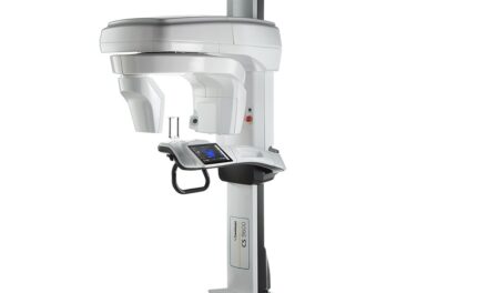
The benefits of cone beam technology are well documented. This technology provides detailed 3D images of patients’ entire jaw and skull structures, shortens scan and appointment times, and exposes patients to significantly less radiation than traditional CT scans. 3D scans improve diagnostic accuracy, treatment precision, and patient satisfaction—right in the orthodontist’s office—and allow clinicians to take advantage of other high-tech tools that streamline efficiency and increase productivity (Figure 1).
While the gains are undeniable, in the light of the current economy, many orthodontists still must be convinced of cone beam’s cost-effectiveness.
My partners and I once shared these sentiments but we have become “believers,” thanks to patient reaction and the positive financial impact that this equipment has had on our practice. We started on our journey with a careful eye on the budget. We had the passion and drive to find a way to reap the benefits of cone beam technology without putting our practice in the red, and we have experienced a positive financial impact.
Our solution was to build a cone beam imaging center, which we predicted would generate referral revenue and grow our practice. Several factors had to align to make the imaging center profitable, offset our investment into the cone beam machine, and achieve improved patient care. Our approach and experience has proven to be a valuable case study in how to fund and obtain the greatest benefit from a cone beam investment.
Our Cone Beam Journey
 |
| Figure 1: 3D radiographic scans enhance diagnostic accuracy, treatment precision, and patient satisfaction. |
Here’s some background information: I practice at Orthodontic Specialists of Green Bay, Wis, with three other orthodontists: Kevin J. Wilke, DDS, MS; Iwei M. Huang, DMD, MS; and Lee S. Bialkowski, DDS. We have been serving patients in the Green Bay, Appleton, and Fox Valley areas for more than 30 years, and I’ve been a doctor here for the past 9 years. Our two main practices are in Green Bay, on the east and west sides, with a satellite practice in Oconto Falls, Wis, totaling 33 chairs. We incorporated cone beam 3D (an i-CAT® by Imaging Sciences International) technology in September 2006.
I was introduced to cone beam technology during the 2001 Moyers Symposium at the University of Michigan School of Dentistry, at a lecture by David Hatcher, DDS, MSc, MRCD (C), a radiologist at DDI Imaging Center. Hatcher has extensive knowledge about 3D technology, and continues to educate other orthodontists through various venues such as the ones offered by Imaging Sciences. Besides Hatcher, I continued to learn more from lectures by experts such as James Mah, DDS, MSc, MRCD, DMSc, an orthodontist and associate clinical professor at both the University of Southern California and the University of Nevada Las Vegas. During a 3-day visit at the oral maxillofacial surgery practice of
G. William Arnett, DDS, in Santa Barbara, Calif, I witnessed his practice’s use of the i-CAT firsthand.
Along with the benefits of the technology, we also learned about selecting the best system for us from these well-regarded academics. My partners and I wanted the improved treatment accuracy, planning, and quality for our patients and practice, but we weren’t sure about what the potential financial rewards would be. Would our new system attract more patients and result in better outcomes? We still had to make a strong business case before going full throttle on the road to 3D.
Pooling our resources seemed like a promising option. In 2006, I organized a meeting in Green Bay with my partners and other leading orthodontists, oral surgeons, prosthodontists, periodontists, and restorative specialists from our area. I wanted to gauge their interest in launching a joint venture to build an imaging center, with cone beam technology as the focal point. Mah was our guest speaker.
Investing in an imaging center would allow my partners and me to benefit from cone beam technology, while creating a profitable referral center with sustained profitability.
After hearing the opinions of our colleagues, my partners and I decided we could make it work together. Here’s how we did it.
Making an Imaging Center a Profit Center
Shared investments in technology can increase business efficiency. Our group practice has a tremendous advantage because we can share ownership expenses across four orthodontists. First, we determined exactly what tools and services to invest in, and the cone beam machine was our “must have.”
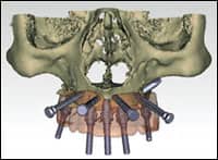 |
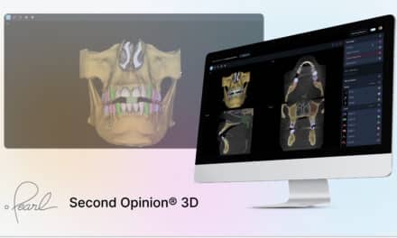
| Figure 2: Active therapeutic application delivered by the imaging center (Image Courtesy of Pi Dental Center). |
We identified what cone beam services we’d offer at the imaging center. From talking to other dentists who would become our referring practitioners, we learned that a primary use of cone beam technology is creating surgical guides such as those used for dental implants.
For this application, the scan data from the cone beam is imported into specialized software that allows us to precisely pinpoint implant locations. A physical guide is then fabricated and acts as a stint during implant-placement surgery (Figure 2).
We recognized we would need to learn this software and learn how to create surgical guides, because many of our external referrals would request this. We also expected to receive referrals from other specialties, such as TMJ dysfunction, pathology, trauma, and sleep apnea. Knowing how to use the machine for a variety of applications improved our cone beam skills for our own patients as well.
To further build revenue and relationships, we hold lectures for our referring dentists to educate them about the technology and our services. We take them through our referral process, and show them how to fill out the Web-based, easy access referral form (Figure 3).
We also try to simplify the payment process by checking and filing patient’s insurance for ourselves and referring dentists and informing patients of their coverage eligibility. Our patients can conveniently pay by cash, check, credit card, or insurance.
Quantifying Return on Investment
Return on investment (ROI) takes into account several unconventional factors, including case
acceptance, treatment quality, treatment success rate, and patient satisfaction. Using our cone beam 3D system every day is crucial in maximizing the benefits.
 |
| Figure 3: The software system we use includes referral tools for colleagues. |
Here’s an example:
We integrated SureSmile® technology with our cone beam capabilities. We’re fortunate that the i-CAT is actually the only cone beam unit that is certified by SureSmile, and proud that we were instrumental in Imaging Sciences’ gaining this certification. Combining these two technologies means that we can utilize the second active therapeutic application developed in dentistry thus far (the first being surgical guides).
SureSmile’s original method of orthodontic movement provides for a new combination of tools, such as 3D imaging and CAD/CAM, that cuts treatment time from 24 months to about 14. The process includes a handheld digital intraoral camera called an OraScanner, which takes hundreds of images of a patient’s teeth and renders them into a 3D model on a computer screen. Orthodontists use SureSmile software to determine teeth’s final position and plan treatment.
Instead of using the OraScanner, we now use the i-CAT to create the 3D CAD model that SureSmile requires. Our i-CAT shortens scan time from 30 minutes to 20 seconds. Since the scan offers full anatomical details, we can see the roots in addition to the crowns of the teeth, and the result is more accurate SureSmile setups. Plus, the radiographic scan can also be used for diagnostic applications (Figure 4).
The time savings is a big part of our ROI. We’ve calculated the value of each clinical minute at our practice at approximately $5. Using the i-CAT instead of the OraScanner, we typically shorten an appointment time by at least 30 minutes; that’s $150 in savings. For 1,000 possible new appointments for braces yearly, that’s $150,000 in savings.
Shorter appointments also lead to higher productivity. Using cone beam 3D scans, we see the same number of patients, about 75 to 125 per day depending on my clinical schedule, in a 3-day clinical week that we used to in a 4-day week, which reduces our staff requirements.
While our referral revenue continues to contribute to our ROI, we now attribute a significant portion of our revenue to the frequent use of our cone beam machine for diagnostics, records, and our SureSmile scans in our practice. In the first
2 full years we had the cone beam and imaging center, we generated $45,000 in additional revenue due to external i-CAT referrals. If our pace continues, we’re projected to generate approximately $110,000 in additional revenue after 5 years of having 3D radiography and launching the imaging center.
Our Cone Beam Buying Guide
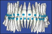 |
| Figure 4: 3D CBCT radiography as seen in SureSmile. |
Our success is also attributable to the fact that we chose a model that fits our practice, workflow, and budget. The wide range of machines on the market offer different features, functionalities, software compatibilities, user-friendliness, and varying price points.
Fortunately, around the time we decided to buy a machine, my partners and I attended the Chicago Dental Society Midwinter Meeting. Talking to cone beam vendors, watching their product demos, and seeing their machines were instrumental to our decision-making process.
The following tips have enabled us to make cone beam a cost-effective investment for our practice:
- Know the company’s leadership. At the Midwinter Meeting, we talked to CEOs and CTOs to learn more about their company’s history, technology, and mission. It’s important to choose a company that’s committed to research and development, because technology changes every day. Look for a company that continues to innovate and improve its products at the speed of industry developments.
- Interview the IT and support team. You want the company to be able to quickly answer your questions and troubleshoot or repair your machine. Imaging Sciences, for instance, guarantees their technical support team will respond to inquiries within 24 hours. If the problem can’t be solved over the phone, the company provides a local network of certified technicians that responds on-site within 48 hours. Other vendors I’ve dealt with over the years have similar policies, but they haven’t always kept their promises.
- Look for the shortest reconstruction times. The time it takes to display a scanned image on the computer screen varies among machines. The longest I’ve seen is several minutes. The shortest I’ve seen is 20 seconds, provided by i-CAT. Lengthy reconstruction times counteract the cost-effectiveness of your cone beam investment. The faster you can view images, the quicker you can begin diagnosis, treatment, and care (Figure 5).
- Talk to cone beam users. We learned a great deal of information when we attended conferences and lectures by other orthodontists who are cone beam owners. This peer education was invaluable, because we obtained objective evaluations and recommendations for what to look for in a cone beam machine.
Spreading the Word
 |
| Figure 5: CBCT has the capability for rapid positioning, scan, and reconstruction times. |
Because of my positive experiences I’m now guiding others on the journey to cone beam ownership. I recently presented lectures at both the University of Minnesota and Marquette University, and showed the students four images taken using a 2D machine. Then I showed them those same images taken with cone beam. The difference was dramatic.
Students clearly understood and accurately identified various dental issues in the 3D images. Cone beam really opens a whole new world to orthodontists. Moving from 2D to 3D imaging is like going from dial-up to high-speed Internet. My infatuation with the technology initially motivated me to find a way to bring it into our practice. By thinking creatively and tapping into our knowledge network, we made the cost-effectiveness apparent.
|
To read more on this subject, search for “” in our archives. |
We think our approach is a great way for orthodontists to integrate cone beam technology into their practices. Thinking outside the box and working with colleagues—even competitors—can be the solution to adding cone beam technology to your practice and patients.
Edward Y. Lin, DDS, MS, maintains a full-time private group practice in Green Bay, Wis. He has lectured extensively and has also taught at both Marquette University and the University of Minnesota Orthodontic Residency Programs. He was a speaker at the International Congress on 3D Dental Imaging in 2008, and will be a returning presenter for the 3rd Congress in June 2009. He can be reached at