by Dipak N. Chudasama, BDS, MSC, MORTH RCS, MBA, and Laurance Jerrold, DDS, JD
Using BioMers translucent archwire
 |
 |
| Dipak N. Chudasama, BDS, MSC, MORTH RCS, MBA | Laurance Jerrold, DDS, JD |
The BioMers archwire was introduced into the orthodontic market at the 2008 AAO Annual Session. This release followed successful clinical trials at two American postgraduate orthodontic programs. At the School of Orthodontics at Jacksonville University, 46 patients underwent a pilot study using a 0.018-inch BioMers wire for up to a 4-month period. The focus of the study was the initial phase of therapy dealing with leveling, aligning, and rotation. Specific inclusion criteria were used, and the entire protocol was approved by the University’s Institutional Review Board. Patients for whom wire deflections with angulations of greater than 60º were necessary were excluded from the study. The results of a pilot study comparing 0.018 BioMers wire to a 0.014 nickel titanium wire over the initial phase showed no differences in the correction of incisor irregularity between the two groups.
The BioMers archwire is made from a polymer composite material, so it fails in a brittle manner, similar to ceramic brackets. Consequently, it was noted that archwire breakages occurred in the premolar/molar area in some patients. Often these patients admitted to eating hard foods such as nuts or candy. It should be emphasized here that 98% of all breakages occurred in the posterior sections of the arch. The anterior sections showed good alignment, and archwire breakage in this segment was a rare occurrence. To minimize posterior breakages, the principal investigators used several clinical modifications.
Clinical Modifications
- We inserted the BioMers wire into plastic tubing, which was then inserted into the bracket slot in the premolar section. The plastic tubing was not engaged into the molar tube (Figure 1). The inner tube diameter has to be greater than 0.018 in dimension to enable the wire to be inserted, and the tube should be inserted into the bracket slot. It is not possible to engage the tube into the molar slot because the thickness of the tube is greater than 0.028. The plastic tubes can also be used for preventing breakage when large unsupported spans are present, or to minimize stress and, hence, breakage on a wire that undergoes crazing in areas of large deflections. The BioMers wire slides easily into the tube, but appreciable friction would still result if space closure is attempted. The purpose of the tube is to support the wire.
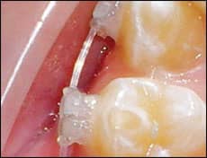
- We placed a metal closed coil around the BioMers wire either in-between the molar and second premolar or the two premolars (Figure 2). The metal coil could also be used to prevent breakage when large unsupported spans of wire lengths are present, but this proved less effective because the metal coils cannot be placed into the bracket slots (unlike the plastic tube). Hence, using the plastic tube is our preference for spans greater than a tooth width. The metal coils are possibly more hygienic than the plastic tubes, so we use them in the shorter interbracket premolar areas when we have areas of wire crazing or fracture.
- A sectional nickel titanium 0.018 archwire can also be used in the lower posterior segment while the BioMers wire is used in the anterior segment. This facilitates maintaining maximal anterior aesthetics while simultaneously correcting molar rotations.
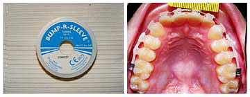 |
| Figure 1: Plastic tubing has been placed on the posterior sections. |
 |
| Figure 2: Metal closed coil was used over a 30-day period, demonstrating that the integrity of the wire was maintained. |
 |
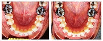
| Figure 3: Full correction of molar rotation has not been achieved. |
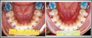
It is interesting that the force levels of an 0.018 BioMers wire were successful at correcting anterior tooth rotations but were not as effective when it came to correcting rotated molars (Figure 3).
Higher force levels are required in the molar areas when rotations are great, and currently our preference is to use sectional wires. With the future introduction of rectangular wires, an intermediate wire would be developed to address this limitation.
The Next Step
 |
| Figure 4. This BioMers wire shows crazing. |
Since the overwhelming majority of wire breakages occurred in the lower premolar/molar area, these measures were employed there. Before a wire actually fractures, an area of crazing is apparent (Figure 4). Crazing does not imply failure of the wire, but once crazing is clinically apparent the measures outlined above should be employed. They can also be used as a preventive measure from initial tooth engagement.
Using the plastic sleeve in the anterior area is usually contraindicated for two reasons. The first is that it negates the esthetic advantage of the wire, and the second is that anterior archwire failures occur in the anterior segment only 2% of the time.
Since the initial study, BioMers LLC has introduced several round archwire dimensions, each having different force levels. This gives the clinician a greater selection of wires to choose from depending on the clinical situation. This will be useful in clinical situations where the incisor irregularity is too severe for a 0.018 wire to be used as the initial leveling arch.
BioMers is currently developing a rectangular archwire. Once this wire passes clinical trials and is coupled with the appropriate bioesthetic bracket system, the orthodontic profession will have a “cradle to grave” aesthetic solution to virtually every orthodontic situation faced in clinical practice.
|
To read more articles by and about composite wire, search our . |
Dipak N. Chudasama, BDS, MSC, MORTH RCS, MBA, is the director of research at the Jacksonville University School of Orthodontics. He can be reached at
Laurance Jerrold, DDS, JD, is the dean and program director at the Jacksonville University School of Orthodontics.