By N.R. Krishnaswamy, BDS, MDS, MOrthoRcs; M. Charles Finny, MDS; and Divyalakshmi Dayalan, MDS
Orthodontic bonding is one of many variables to dictate outcome and efficiency of orthodontic care. A good orthodontic adhesive should keep the bracket bonded to the tooth throughout treatment, avoiding delays, untoward expenses, or patient inconvenience. Also, debonding should not damage the enamel at treatment end.1
Research has concentrated on reliability and effectiveness of adhesive systems and simplifying adhesive technique with many completed prior clinical studies evaluating different brands of adhesive systems.2-4 Nevertheless, few studies have assessed two different adhesive systems in the same patient by using quadrant variations.
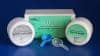
The objectives of this clinical trial were to compare bracket failure rate of two different light cure adhesive materials used in bonding of orthodontic brackets to teeth and evaluate the efficiency of each. The two composite adhesives were Transbond XT from 3M Oral Care and Bracepaste from American Orthodontics.
This double blinded prospective study randomly selected patients to compare bond failure rates over a 10-month period. Bracket failures were analyzed between dental arches, regions, sides, quadrants, age, gender, and archwire composition and dimensions.
Angle Class I or Class II malocclusion patients presenting for orthodontic treatment and willing to participate in the study were recruited using the following inclusion criteria: permanent dentition including incisors, canines, premolars, and first molars; teeth with sound, noncarious buccal enamel; and no previous orthodontic treatment with fixed appliances. Subjects were excluded if they couldn’t provide informed consent, or had craniofacial anomalies, or congenital enamel defects. Eventually, 38 patients participated in a single-center, single-operator, split-mouth randomized controlled trial with a 1:1 allocation ratio with no changes after trial commencement.
Interventions
Patients had maxillary incisors, canines, and premolars bonded with metal brackets (Mini Master Series, American Orthodontics) using Transbond XT on two quadrants and Bracepaste on the other two quadrants. The side allocation was randomized as detailed in Table 1.

Each participant’s dentition was divided into four quadrants. Quadrants were switched opposite each other with different combinations in all patients to avoid operator bias. Bonding surfaces were cleaned using slurry of pumice for 10 seconds, thoroughly rinsed for 20 seconds and air dried completely using an airway syringe. Teeth were isolated using cheek retractors, tongue away, and cotton rolls. Teeth were acid etched using 37% phosphoric acid (ScotchBond etchant, 3M Oral Care) for 30 seconds. After thorough washing and air drying, the surface was checked for the frosty appearance of enamel to ensure proper etching. The bonding agent prescribed for both adhesive systems was applied with a microbrush and light cured for 10 seconds using a 3M Elipar (3M Oral Care). With brackets in appropriate positions from second premolar to second premolar in both the arches, excess adhesive was removed using a hand scaler and each bracket was light cured for 10 seconds using 3M Elipar LED. Molars were banded and archwires were placed 10 minutes after bonding.
Adhesive Remnant Index (ARI) was scored when brackets failed by analyzing the bracket base at 10x magnification. Most studies score ARI based on Artun and Bergland’s classification.34 However, a modified 5 scale classification by Bishara and Truelove35 provided better quantitative and qualitative assessment of ARI36 and is more illustrative to describe remnants on failed brackets for this study.
Bracket failures were recorded at each 4-week interval appointment, or on call by patients noticing failures between appointments. Bracket failures were analyzed between the dental arches, quadrants, regions, age, gender, archwire type, and composition.

Results
Bonding occurred on 560 teeth—280 teeth with Transbond XT and 280 teeth with Bracepaste. Bond failures totaled 44 (7.9%), with Transbond XT exhibiting 25 (4.5%) and Bracepaste exhibiting 19 (3.4%). No statistically significant difference between bracket failures with Transbond and Bracepaste was noted (p value = 0.346) (see Table 2).

Between arches, 25 bond failures existed in the mandible and 19 in the maxilla. The second premolars (22 brackets) had the highest bond failures. Between quadrants, 24 right side bond failures resulted with 20 left side. More bond failures presented in patients older than 18 and males experienced higher rates than females. In both groups, greater bond failure occurred with round archwire than rectangular archwire and more with NiTi wires than stainless.
ARI displayed a highest score of 5 in 37 teeth depicting 100% of adhesive at the bracket base. Seven brackets scored 4, denoting more than 90% of adhesive on the bracket base. Transbond XT scored 4 in three instances of bond failure and scored 5 in 22 instances. Bracepaste scored 4 in four instances of bond failure and scored 5 in 15 instances.
Discussion
Previous studies tested effectiveness of adhesive materials in vitro.5-7 In vitro studies have not been successful predicting in vivo effectiveness and very few studies have been conducted in vivo.8,9
Failure rate is a common clinical performance indication of bonding adhesives, allowing natural comparisons between studies.10 Therefore, this study was an in vivo split mouth design study (SMDS) to reduce probability of inter-patient variability bias and record bracket failure numbers to analyze and discover the better adhesive system.
In this study, 44 brackets (7.9%) exhibited bond failures among 560 bonded teeth, within the range of failure percentages (0.5% to 16% with an average of 6%) reported in previous in vivo studies.11-13
Patients older than 18 demonstrated higher failure rate. In late adolescents and adults, the occlusal forces—mainly bite or chewing force—are reported as an important factor in bond failure. Various studies show bite force increases proportionately with age due to developing muscle forces between 6 and 18 years of age and decreases after 25 years in females and 45 years in males. This factor possibly led to higher number of failures in individuals above 16 to 18 years.14,15
Gender variations on failure rates show males having a greater failure percentage than females.16,17 Few studies displayed females to have insignificant but higher failures, which were substantiated by the number of increased sample size.18-20 In this study, higher failure rate was noted in more males than females. A statistical significance of p value < 0.5 denoted a predominance of males. Nikolaos Koupis17 and Imad Shamma21 claimed females are more attentive and better maintain fixed appliances. Predominance in bracket failures in males was also attributed to the increased bite forces.22
Within the arch, the mandibular displayed a higher number of clinical bracket failures than maxillary but not of statistical significance. Past literature studies show a significant trend with mandible showing higher failure rate than maxilla despite the various techniques, type of material, or light curing unit used.19,22,23 Previous studies substantiate high occlusal forces as the common factor in predominant mandibular arch failures.24
No statistical significance between the sides existed between the two adhesives. Literature documents two broad notions in elucidating the failure between different segments. First, the clinician’s right-handedness or left-handedness plays a vital role in bracket bonding efficiency within the segments. A right-handed clinician tends to show ease in bonding technique on the right segment of the patient, especially due to better visibility, moisture control, and positioning of brackets.24,25 However, Kinch et al26 claimed a right-handed clinician found better access in bracket placement on the left side. Second, the patient’s habitual manner to chew food on a particular side, and inequality in the forces applied during brushing, may cause archwire distortion, leading to bracket failure.26
A statistically significant value existed with posterior teeth with the mandibular second premolar displaying the maximum bond failure rate. Contaminations due to either saliva, gingival fluid seepage, or blood may lead to reduced bond strength. The proximity of salivary duct opening causes difficulty in controlling the salivary flow in posterior segment.
Heavy occlusal forces or masticatory forces and, most importantly, the maximum voluntary bite force or chewing force are higher in the posterior region of dentition.27 The mandibular anterior teeth, though highly predisposed by the overbite, report less failures due to decreased incisal bite force.28 Visibility and accessibility reduced from anterior to posterior part of the dentition leading to bracket positioning difficulty.18,23 Uneven resin thickness distribution was found in failed brackets of posterior teeth. Knoll29 described it as a significant reason for bond failure. Thicker adhesive areas may increase polymerization shrinkage and differences in coefficient of thermal expansion/contraction, thus building stresses within the resin resulting in bond failure.29,30
Kusy et al31 found that no ideal archwire exists, since the demands of each treatment stage necessitate different characteristics emphasizing the stress/strength ratio. In this study, NiTi wires displayed highest bond failure rates. This may be attributed to the initial treatment stages where teeth are significantly malpositioned and more vulnerable to cuspal contacts from opposing teeth.
Several authors note failure mostly occurs at the bracket-adhesive interface.32,33 However, this study correlates with studies by Sfondrini et al34,35 and Henkin et al36 claiming bond failure occurred more predominantly at the enamel-adhesive interface. Enamel interface breakage signifies poor efficiency of adhesive as chemical bond and partial mechanical bond created by penetration of the adhesive on the etched enamel surface is vital. The enamel surface bond is technique sensitive and depth of etch shortcomings may lead to a weak bond.32
The survival rates plotted revealed most bracket failures occurred within the first 3 months. Fifty percent of bracket failures occurred within the first 65 days with Transbond XT and 62 days for Bracepaste, similar results to previous studies showing initial 3 to 6 months portraying higher failure rates.37,38
An enamel surface failure can be considered beneficial. It may demonstrate that bracket mesh, which also plays an important role in bonding, has a satisfactory bond strength and the bracket adhesive interface bond strength is higher than that of the enamel adhesive interface. Only seven brackets showed cohesive failure with the majority showing adhesive failure. Thus, it can be safely assumed in this study that the efficiency of the two adhesive materials is optimal for use in practice.
Conclusion
The following conclusions were drawn from this study:
- Both light cure composite resin adhesive materials, Transbond XT and BracePaste, display adequate and optimal bond strength suitable for application in orthodontic bonding procedures in day-to-day clinical practice.
- Age differences did not statistically affect the failure rate; however, males demarcated significantly higher failures.
- Within the dentition, posterior teeth had significantly higher failure rate. The overall differences in quadrants and arches depicted higher failures in right quadrant and mandibular arch, but did not reach a statistically significant threshold.
- Following ideal protocols is advised in allied procedures and techniques such as isolation, etching, curing, etc, which play major roles in bracket-adhesive-enamel interface. OP
N.R. Krishnaswamy, BDS, MDS, MOrthoRcs, is a professor and head of the department of orthodontics and the vice principal of Ragas Dental College & Hospital, Chennai, India. A graduate of Madras Dental College, he obtained his Masters in Orthodontics from College of Dental Surgery, Manipal. A fellow of the Royal College of Surgeons of Edinburgh and a diplomate of the Indian Board of Orthodontics and The National Board of Medical Sciences, he received the AAO’s B.F and Helen E. Dewel Clinical Research Award and the Japan Dental Association’s International Scientific exchange fund. Krishnaswamy served as the president of the Indian Orthodontic Society and chairman of the Indian Board of Orthodontics.
Additional Authors: M. Charles Finny, MDS, orthodontist, Chennai, and former graduate student in the department of orthodontics at Ragas Dental College and Hospital; and Divyalakshmi Dayalan, MDS, senior lecturer in the department of orthodontics, Ragas Dental College & Hospital, Chennai
References
- Bakhadher W, Halawany H, Talic N, Abraham N, Jacob V. Factors Affecting the Shear Bond Strength of Orthodontic Brackets – a Review of In Vitro Studies. Acta Medica (Hradec Kralove, Czech Republic). 2015;58(2):43–8.
- Lovius B.B.J, Pender N, Hewage S, O’Dowling, Tomkins A, A Clinical Trial of a Light Activated Bonding Material over an 18 month Period, Br J Orthod, 1987; 14:1; 11-20.
- Elekdag-Turk S, Isci D, Turk T, Cakmak F. Six-month bracket failure rate evaluation of a self-etching primer. Eur J Orthod. 2008 Jan 21;30(2):211–6.
- Hegarty DJ, Macfarlane TV. In vivo bracket retention comparison of a resin-modified glass ionomer cement and a resin-based bracket adhesive system after a year. Am J Orthod Dentofacial Orthop. 2002;121:496–501.
- Alexandre P, Young J, Sandrik JL, Bowman D. Bond strength of three orthodontic adhesives. American Journal of Orthodontics and Dentofacial Orthopedics. 1981;79(6):653–660.
- Bishara SE, Olsen ME, Jakobsen JR. Evaluation of a new light-cured orthodontic bonding adhesive. Am J Orthod Dentofacial Orthop. 1998;114(1):80–87.
- Trimpeneers LM, Dermaut LR. A clinical trial comparing the failure rates of two orthodontic bonding systems. Am J Dentofac Orthop. 1996; 110:547-50
- Finnema KJ, Özcan M, Post WJ, Ren Y, Dijkstra PU. In-vitro orthodontic bond strength testing: A systematic review and meta-analysis. Am J Orthod Dentofacial Orthop. 2010 May;137(5):615–622.e3.
- Verma SK, Maheshwari S, Tariq M, Khan S. The inadequacy of in-vitro orthodontic bond strength testing in clinical application. Intl J Dent Sci Res. 2013 May;1(2):54–7.
- Cozza P, Martucci L, De Toffol L, Penco SI. Shear bond strength of metal brackets on enamel. Angle Orthod. 2006;76(5):851–856.
- Hobson RS, McCabe JF, Rugg-Gunn AJ. The relationship between acid-etch patterns and bond survival in vivo. Am J Orthod Dentofacial Orthop. 2002 May;121(5):502–9.
- Sunna S, Rock WP. Clinical performance of orthodontic brackets and adhesive systems: a randomized clinical trial. Br J Orthod 1998;25:283-7
- Zachrisson BU. A posttreatment evaluation of direct bonding in orthodontics. Am J Orthod Dentofacial Orthop. 1977;71(2):173–189
- Bakke M, Holm B, Jensen BL, Michler L, Moller E. Unilateral, isometric bite force in 8–68 year old women and men related to occlusal factors. Scand J Dent Res. 1990;98:149–158.
- Goyal A, Hurkadle J, Magegowda S, Bhatia P. Use of light-curing units in orthodontics. J Inv Clin Dent. 2013 Aug;4(3):137–41.
- Verma SK, Maheshwari S, Tariq M, Khan S. The inadequacy of in-vitro orthodontic bond strength testing in clinical application. Intl J Dent Sci Res. 2013 May;1(2):54–7.
- Koupis NS, Eliades T, Athanasiou AE. Clinical Evaluation of Bracket Bonding Using Two Different Polymerization Sources. Angle Orthod. 2008 Sep;78(5):922–5.
- Cal-Neto JP e, Quintão CA, de Oliveira Almeida MA, Miguel JAM. Bond failure rates with a self-etching primer: A randomized controlled trial. Am J Orthod Dentofacial Orthop. 2009 Jun;135(6):782–6.
- Linklater RA, Gordon PH. Bond failure patterns in vivo. Am J Orthod Dentofacial Orthop. 2003;123(5):534–539.
- Thind BS, Stirrups DR, Hewage S. Bond failure of gingivally offset mandibular premolar brackets: A randomized controlled clinical trial. Am J Orthod Dentofacial Orthop. 2009 Jan;135(1):49–53
- Shammaa I, Ngan P, Kim H, Kao E, Glawdin M, Gunel E, BrownC. Comparison of bracket debonding force between two conventional resin adhesives and a resin-reinforced glass ionomer cement: An invitro an in vivo study. Angle Orthod 1999;69:463-469.
- Pandis N, Eliades T. A comparative in vivo assessment of the long-term failure rate of 2 self-etching primers. Am J Orthod Dentofacial Orthop. 2005 Jul;128(1):96–8.
- Krishnaswamy NR, Sunitha C. Light-emitting diode vs halogen light curing of orthodontic brackets: A 15-month clinical study of bond failures. Am J Orthod Dentofacial Orthop. 2007 Oct;132(4):518–23.
- Adolfsson U, Larsson E, Ögaard B. Bond failure of a no-mix adhesive during orthodontic treatment. American journal of orthodontics and dentofacial orthopedics. 2002;122(3):277–281.
- Sunna S, Rock WP. Clinical performance of orthodontic brackets and adhesive systems: a randomized clinical trial. Br J Orthod 1998;25:283-7
- Kinch AP, Taylor H, Warltler R, Oliver RG, Newcombe RG. A clinical trial comparing the failure rates of directly bonded brackets using etch times of 15 or 60 seconds. Am J Orthod Dentofacial Orthop. 1988;94(6):476–483
- Oz AA, Oz AZ, Arici S. In-vitro bond strengths and clinical failure rates of metal brackets bonded with different light-emitting diode units and curing times. Am J Orthod Dentofacial Orthop. 2016 Feb;149(2):212–6.
- Cozza P, Martucci L, De Toffol L, Penco SI. Shear bond strength of metal brackets on enamel. Angle Orthod. 2006;76(5):851–856
- Knoll M, Gwinnett AJ, Wolff MS. Shear strength of brackets bonded to anterior and posterior teeth. Am J Orthod Dentofacial Orthop 1986; 89:476-479
- Galindo HRA, Sadowsky PL, Vlachos C, Jacobson A, Wallace D. An in vivo comparison between a visible light-cured bonding system and a chemically cured bonding system. Am J Orthod Dentofacial Orthop. 1998;113(3):271–275
- Kusy RP. A review of contemporary archwires: their properties and characteristics. Angle Orthod.1997;67(3):197-208.
- Artun J, Bergland S. Clinical trials with crystal growth conditioning as an alternative to acid-etch enamel pretreatment. Am J Orthod Dentofacial Orthop. 1984;85(4):333–340.
- Pinto CM de S, Ferreira JTL, Matsumoto MAN, Borsatto MC, Silva RAB da, Romano FL. Evaluation of different LED light-curing devices for bonding metallic orthodontic brackets. Braz dent J. 2011;22(3):249–253
- Sfondrini MF, Cacciafesta V, Scribante A, Boehme A, Jost-Brinkmann P-G. Effect of light-tip distance on the shear bond strengths of resin-modified glass ionomer cured with high-intensity halogen, light-emitting diode, and plasma arc lights. Am J Orthod Dentofacial Orthop. 2006 Apr;129(4):541–6.
- Sfondrini MF, Cacciafesta V, Scribante A, De Angelis M, Klersy C. Effect of blood contamination on shear bond strength of brackets bonded with conventional and self-etching primers. Am J Orthod Dentofacial Orthop. 2004 Mar;125(3):357–60.
- Henkin F de S, Macêdo É de OD de, Santos K da S, Schwarzbach M, Samuel SMW, Mundstock KS. In vitro analysis of shear bond strength and adhesive remnant index of different metal brackets. Dent Press J Orthod. 2016 Dec;21(6):67–73.
- Elekdag-Turk S, Isci D, Turk T, Cakmak F. Six-month bracket failure rate evaluation of a self-etching primer. Eur J Orthod. 2008 Jan 21;30(2):211–6.
- Hegarty DJ, Macfarlane TV. In vivo bracket retention comparison of a resin-modified glass ionomer cement and a resin-based bracket adhesive system after a year. Am J Orthod Dentofacial Orthop. 2002;121:496–501.