The new guidelines emphasize clinical necessity before ordering radiographs and address specific uses in orthodontics, including panoramic imaging.
The American Dental Association has published new recommendations for ordering dental imaging, advising that radiographs should only be used when clinically necessary to support diagnosis and treatment planning.
The guidelines, which update recommendations from 2012, address both planar dental radiography and cone-beam computed tomography. They emphasize the importance of conducting a clinical exam before taking images to determine if they are needed based on an individual patient’s history and clinical findings.
Focus on Clinical Necessity
The recommendations were developed by an expert panel from the ADA Council on Scientific Affairs and are endorsed by the American Academy of Oral and Maxillofacial Radiology.
“Dental imaging is an important diagnostic tool that can help improve oral and overall health outcomes when used appropriately,” says Erika Benavides, DDS, PhD, lead author and clinical professor at the University of Michigan School of Dentistry. “Similarly, dental X-rays should be ordered only after first examining the patient’s medical and dental histories, prior X-ray images and current clinical exam findings.”
Orthodontic-Specific Guidelines
The report includes recommendations for various specialty areas, including orthodontics. For orthodontic applications, the panel advises:
- Using panoramic radiographs as the initial imaging modality for monitoring tooth eruption before initiating treatment.
- Using panoramic radiographs to assess root alignment during treatment.
- Prescribing radiographs judiciously and using effective dose-reduction methods for children and young adults.
- Using panoramic radiography for assessment and treatment planning when there is a clinical indication for radiographic evaluation of third molars, supernumerary, and supplemental teeth.
- Considering panoramic radiography for initial imaging of patients with suspected temporomandibular joint disorders to rule out gross osseous abnormalities, though its low sensitivity may mean it is not sufficient for a definitive diagnosis.
READ MORE: Why CBCT Is Key to Long-Term Case and Professional Stability
Additional Clinical Recommendations
The guidelines also cover other clinical scenarios, including patient appointment types and risk factors for dental caries or periodontal disease. For periodontal disease management, the panel notes that a 2D full-mouth series combined with a clinical exam remains the standard for evaluation. CBCT is not supported for periodontal disease management except for treatment planning in complex cases.
The panel also recommends panoramic radiography for initial assessment before dental implant procedures, with CBCT used for presurgical planning and placement.
This report is the second in a two-part series on optimizing diagnostic imaging in dentistry. The first installment, published in JADA in 2024, focused on radiation safety and regulatory matters, including the recommendation to discontinue routine thyroid and abdominal shielding—a move that built upon 2023 guidance from the American Academy of Oral and Maxillofacial Radiology.
“Dental X-rays are safe. In some cases, a dental X-ray delivers less radiation than a single day of the natural radiation we are all exposed to just by living in the world,” Benavides said. “Yet it is important to follow the ALARA (As Low As Reasonably Achievable) principle recognized in both dentistry and medicine to minimize exposure over a patient’s lifetime and only order X-rays when clinically necessary,” Dr Benavides says.
The full recommendations were published in the January issue of The Journal of the American Dental Association.
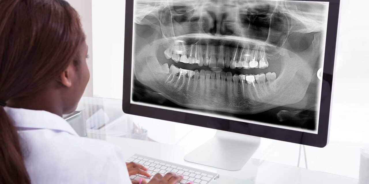
Photo: ID 40192946 © Andrey Popov | Dreamstime.com