by Michael C. Alpern, DDS, MS; and Douglas G. Nuelle, MD, FACS
Records, referrals, and initial treatment
 |
This is Part 2 of a two-part approach to TMJ symptoms, or TMD. The reader may appreciate this part if he or she first reads Part 1 (from last month’s issue). This article will discuss diagnostic records and referrals as well as initial conservative treatment.
Diagnostic Records and Referrals
After taking a complete medical and dental history and doing the clinical examination and analysis mentioned in Part 1, the orthodontists should take selective diagnostic records. I wrote my orthodontic master’s thesis on panoramic radiography. So, my first generalized scanning is to take a unique panoramic digital radiograph. Please bear in mind that there are a myriad of panoramic x-ray machines. I chose the Planmeca ProMax because this particular unit has three axes of rotation, enabling the x-ray focal trough to be individualized to each patient’s anatomy. This increases the accuracy of the image, especially in the TMJ areas. I believe the choice of a panoramic machine should include an adjustable focal trough. Many generic panoramic machines lack an adjustable focal trough, so the TMJ image may be distorted horizontally, vertically, and angularly. However, even the best machines must be operated by a dentist, hygienist, trained assistant, or orthodontist who has experience in setting the correct focal trough as accurately as possible.
After taking the panoramic image, the clinician should carefully examine it from one TMJ to the other. I look at all teeth and their surrounding periodontium. Naturally, I scan for tumors, cysts, abscesses, or other abnormalities. I look for any teeth with large, deep questionable restorations, missing teeth, drifted or malpositioned teeth—anything that could be causing pain or dysfunction (especially any encumbrance to freedom of mandibular motion).
 |
| Michael C. Alpern, DDS, MS |
 |
| Douglas G. Nuelle, MD, FACS |
I also make a lateral cephalometric radiograph taken in Frankfurt Horizontal position. I digitize this image and make multiple measurements from differing author’s opinions. In particular, I look for potential loss of vertical support that could abnormally load the fragile TMJ fibrocartilage covering the entire articular fossae and the TMJ disks.
Based on these images and everything gleaned from Part 1, I may then refer the patient for full-mouth periapical radiographs and evaluation by a periodontist, endodontist, oral surgeon, ENT, and/or physicians specializing in sleep apnea disorders. In addition, every TMD patient is referred to a PhD clinical neuropsychologist and a board-certified neurologist.
The PhD clinical neuropsychologist must be trained in TMJ evaluations. This doctor has the patient complete an Minnesota Multiphasic Personality Inventory (MMPI), a Strait, Trait Anger examination (STAXI), and a psychological examination. These three tests focus on all factors that may be causing the patient stress, potentially leading to abnormal internalizing of their daily and previous cumulative stress. This, in turn, may cause the patient to exhibit this stress in any number of destructive habits that can cause abnormal loading of the fragile TMJ fibrocartilage. Please remember from Part 1 that human fibrocartilage has no nerve supply, so it cannot inform the patient of the permanent damage occurring. Worse, human fibrocartilage lacks any blood supply, making normal healing nearly impossible.
The clinical neuropsychologist is charged with the diagnosis of all primary stress components leading to destructive habits and begins (with the patient’s consent and cooperation) destructive habit-control training.
For communication purposes, the dentist or orthodontist is informed of the psychologist’s findings and treatment using the Wharton TMJ rating system.1 This rating ranges from 1 to 5. A 1 rating infers minimal stress, good cooperation, and good anticipated destructive habit-control training. A 5 rating indicates severe uncontrolled stress and destructive habits. For a 25-year unpublished study I am conducting, I treat only patients rated 1, 2, and 3. I have experienced a 100% failure rate attempting to treat 4s or 5s. Thus, all 4s and 5s are informed that stress and destructive habits are the major component in their TMD problem and it is not safe to attempt any other treatment until they improve their destructive habit-control training and stress control by working with a psychologist and possibly a psychiatrist until their rating is at least a 3.
In 33 years of seeing TMD patients, I have found that every single one of these patients lean on their jaws with their hands or fists. When I mention to a patients that they lean on their jaw, they always deny it. Yet, consistently during the rest of the examination, whether speaking or listening, these same patients constantly lean on their jaw. This constantly loads the TMJ fibrocartilage and destroys it.
 |
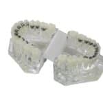
| Mounted models have an aperture created by opening the front pin of the polycentric hinge joint articulator to vertically unload the TMJs. |
Orthodontists are not trained in destructive habit-control training. A few clinical psychologists are trained in this therapy. You may have to search your community to find a psychologist willing to study my textbook and research destructive habit-control training. Without this component in your arsenal of treatment, you may experience a high degree of failure.
The second most critical component in TMJ DJD diagnosis is referral to a board-certified neurologist. This is especially important if the patient has a history of awakening with a headache two to three times per month. These patients must be evaluated by a board-certified neurologist because there are many serious, even life-threatening, problems that can mimic TMJ symptoms in the head.
One memorable patient came to our team years ago. We were sent a patient for a second opinion. She was scheduled to have both TMJs replaced and have a simultaneous maxillary three-piece LaForte I and mandibular advancement. Our neurologist found that she had left temporal lobe epilepsy. She was placed on a small amount of Dilantin and all appliances were removed. Unfortunately, I have seen TMJ patients for second opinions who came into my office having worn splints for several years. One had an aneurysm pulsing up through the back of her throat. Two others had brain tumors.
Our neurologist is charged with giving each patient a complete medical and neurological clearance. He is also charged with all pain control. A significant number of patients presenting with TMJ symptoms were found to have undiagnosed prolapsed mitral valves. TMD is a disease of the viscoelastic tissue, as is prolapsed mitral valve.
Initial Conservative Treatment
After clearance and/or classification by the psychologist and the neurologist, if the patient has pain and or dysfunction in one or both TMJs, we refer the patient for TMJ imaging. We define dysfunction as difficulty in chewing, talking, or swallowing. Technology is changing TMJ imaging. We currently use corrected parasaggittal tomographic images. These are programmed from a submentovertex cephalometric radiograph. We carefully measure the inter-condylar angles and distances from the center of odontoid. We use 1-mm cuts from the lateral pole to the medial pole of each condyle.
 |
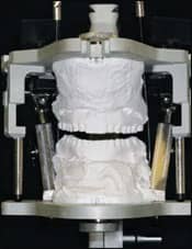
| Maxillary and mandibular full-arch, flat-plane splints are made to accommodate the amount of vertical unloading required. |
Should any of these appear abnormal, we then do a CT scan for 3D reconstructive imaging—although my favorite is cine 3D MRI. These images show the movement of the condyles and the TMJ disks so that we can judge if they move together without any encumbrances.
Based on all the diagnostic information, we begin with a vertically unloading, full-arch, flat-plane maxillary bite splint constructed on a programmed polycentric hinge joint patient articulator. We program vertical opening in to produce flat-plane splints requiring little to no adjustment. Patients are required to wear TMD splints full-time, including while they are eating. We treat patients as if they were recovering from orthopedic surgery: If you sprained or fractured your ankle, and your doctor placed you in a cast, you would not remove the cast three or four times a day and run on your injured ankle and expect the ankle to heal. Thus, full-time wear is required. Our splints are constructed with a new Glenroe-Essix material, which is very clear and has been very acceptable to patients.
Combining this treatment with the psychologist controlling destructive habits and the neurologist controlling clenching during REM sleep, our success rate over the last 25 years has been approximately 95%.
In an initial unpublished study, my neurologist has been titrating Klonopin taken prior to sleep. The dosage is individualized for each patient. I have monitored the clenching grooves in patients on Klonopin and found a significant decrease in abnormal parafunctional mandibular grinding grooves in their splints. It has been my experience that patients on neurologist-prescribed Klonopin and full-time splint therapy have the best success rate in conservatively relieving TMJ symptoms.
After the splints are placed, we see the patient in 1 week, then 3 weeks, then every 8 weeks. If the patient finds relief of some symptoms, but not clicking or clunking, then the casts are remounted on a programmed POLY articulator, which reproduces all the patient’s chewing strokes in the laboratory, and the front pin is opened between 4 to 6 mm. Appropriate acrylic is added to the splint and “chewed in.” Thus, reseating a splint with increased vertical unloading requires only a few minutes until all teeth contact.
I have attempted to describe (in a brief article) a conservative method of obtaining diagnostic records and referrals and then placing a full-arch maxillary flat-plane splint.
This is an extensive subject requiring more than this article permits. If you require additional information, please contact me at the e-mail address below.
Michael C. Alpern, DDS, MS, is in private practice in Port Charlotte, Fla. He can be reached at
Douglas G. Nuelle, MD, FACS, is in private practice in orthopedic surgery in Blue Mountain, Ga. He is the author of numerous lectures and articles on TMJ arthroscopy. He can be reached at
Reference
- Alpern MC. The OrthoEvolution: the Science and Principles behind Fixed/Functional/Splint Orthodontics. Bohemia, NY: GAC International; 2003:91-100.