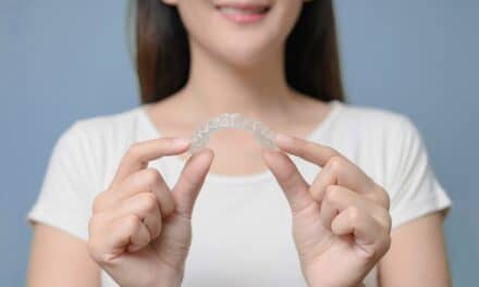
A look at how the Invisalign Specifix Attachment System supports root control, aligner engagement, and treatment efficiency while simplifying attachment placement and removal.
By Manal Ibrahim LaVacca, DDS
Attachments are commonly used during clear aligner orthodontic treatment to improve control of complex tooth movements such as anterior extrusion, posterior intrusion for open-bite correction, canine rotation, and root uprighting during space closure or implant site development. Common issues associated with composite attachments formed using a thermoformed attachment template include overfill, underfill, voids, and formation of composite flash. Composite attachments also require careful shade matching for optimal esthetics and may chip, stain, or wear down during treatment, which can compromise their appearance and performance. For many patients, visible attachments on the anterior teeth can also reduce the esthetic appeal that makes clear aligner therapy attractive.
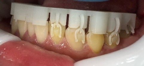
The Invisalign Specifix Attachment System is a 3D-printed attachment solution that uses attachment positioners to accurately place attachments to the precise shape, size, and accurate location per the doctor’s digital ClinCheck treatment plan. This delivery mechanism eliminates the need to fill an attachment template with composite, so aligner adaptation issues related to template overfill and underfill are no longer a problem.
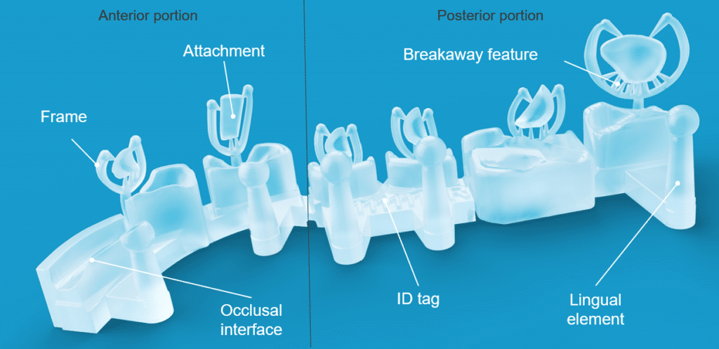
The Specifix attachment material is esthetic and designed to optimize aligner engagement while blending in with the underlying tooth shade. The result is a favorable balance between function and appearance for patients who require attachments on their teeth during their Invisalign treatment. In our experience using Specifix attachments, the clinical workflow has been streamlined, requiring fewer steps and simplifying treatment process.
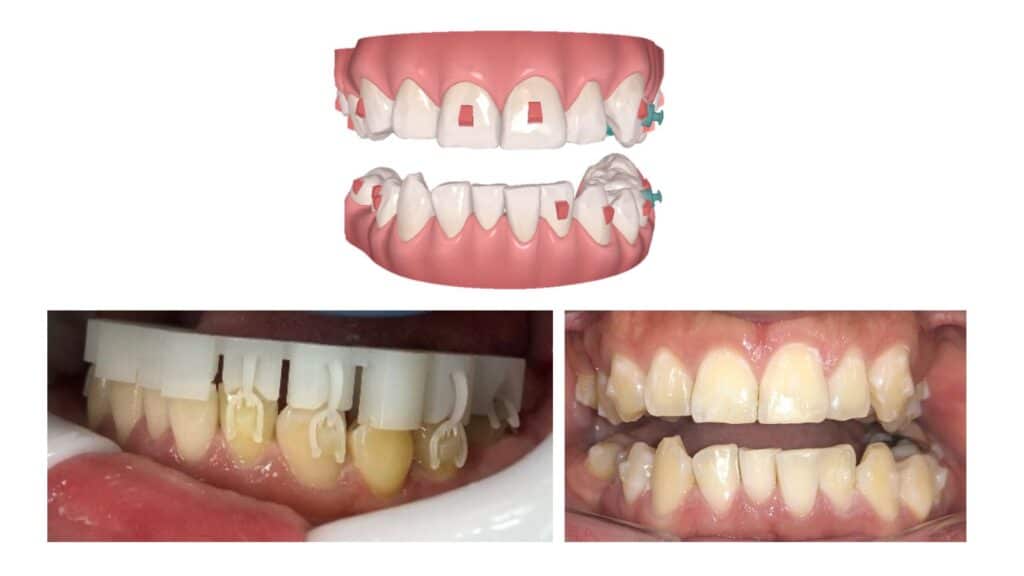
Having treated more than 6,000 clear-aligner patients across a wide range of case complexity, our practice is very familiar with the challenges associated with bonding and removing conventional composite attachments. As an educator and advisor for Align Technology Inc, I have treated hundreds of Invisalign cases using the Invisalign Specifix attachment system during the technical design assessment.
Case 1
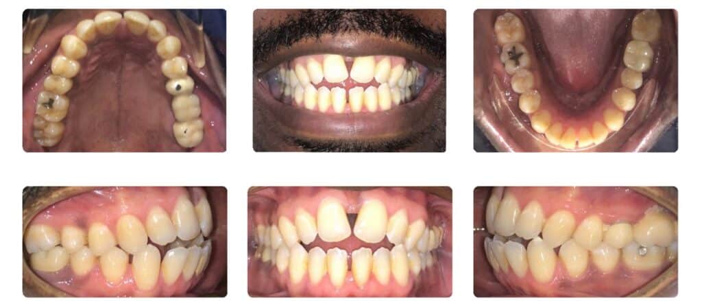
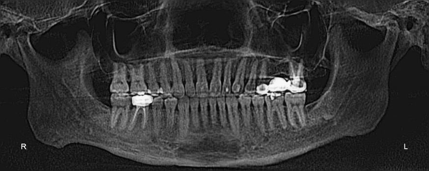
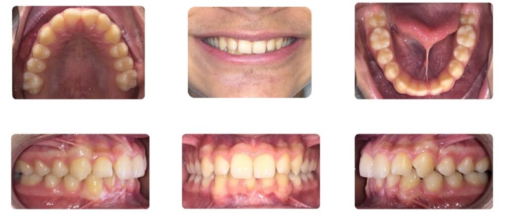
The chief concern of this 49-year, 1-month-old male patient was a maxillary diastema and an anterior open bite. He presented with bimaxillary protrusion, right and left Class I canine and molar relationship, a moderate anterior open bite, negative overjet, narrow arch forms, mesially-tipped maxillary central incisors with divergent roots, maxillary midline frenum with midline diastemas in both arches, and a mandibular midline shift to the right.
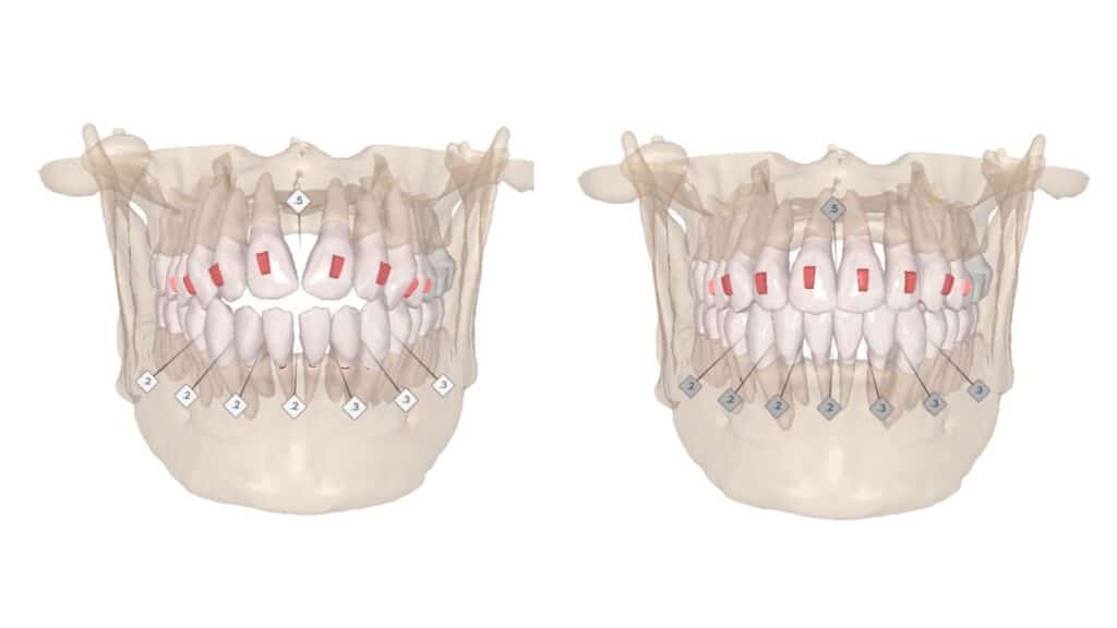
The treatment plan was to close the anterior open bite using Invisalign aligners with vertical rectangular Specifix attachments on U 5-5 to extrude the maxillary incisors and upright the maxillary central incisor roots. Lingual vertical rectangular attachments were prescribed on U 3-3 to provide additional root control of the anterior segment.1 IPR was planned on L 4-4 to retract the mandibular incisors and create positive overjet. IPR between the U1s was planned to broaden the interproximal contact area and help camouflage the deficient interdental papilla.





Case 2
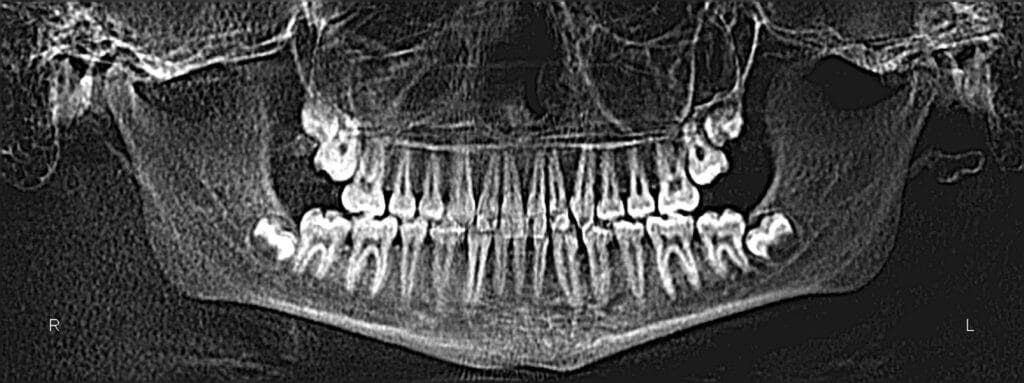
A 13-year, 6-month-old male patient presented with a chief concern of spacing in the lower arch due to a congenitally missing mandibular left lateral incisor. He had a Class I molar and canine relationship on the left and right, a moderate deep overbite, moderate increased overjet, and a mandibular midline shift to the left due to the missing lower incisor.
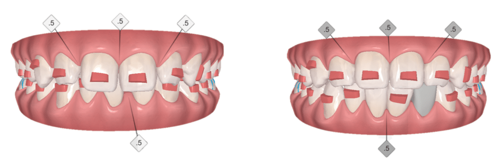
The treatment plan was to idealize space for a resin-bonded bridge and then a dental implant and crown after his growth was completed. To correct the deep bite, the upper and lower incisors would be intruded, so attachments on the canines and premolars were prescribed to prevent vertical disengagement of the aligners during intrusion. IPR on U 2-2 was planned to reduce the anterior overjet. IPR between L1s was planned to optimize the interproximal contact area and reduce the apparent papillary deficiency.
Specifix attachments were prescribed on the lower incisors for root control. Lingual vertical rectangular attachments were prescribed on LL1, 3 and UL2, 3 to enhance root control and aligner coupling in the anterior region.1 Aligner cutouts for bonded buttons were prescribed on U4, 5 (lingual) and L6s (buccal) for Class II elastics to improve the overjet.




Discussion
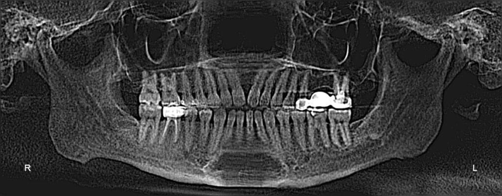
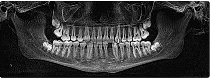
Without good root control of the incisors, the teeth would tend to tip rather than translate. In the two cases presented, the desired root angulation was achieved in the anterior teeth after 12 months of treatment, and healthy root morphology was observed in the progress panoramic radiographs. The planned tooth movements appeared to be fully expressed, as demonstrated by the close correspondence between the clinical outcome and the planned outcome in the ClinCheck treatment simulation. The aligner adaptation during treatment has also been excellent.


In Case 1, the patient successfully underwent a laser frenectomy and is currently completing orthodontic refinement with additional aligners. This phase includes IPR of the maxillary central incisors to maximize the interproximal contact area, plus IPR and retraction of the mandibular incisors to resolve heavy anterior contacts. The attachments were left in place to ensure continued root control of the anterior teeth.
The patient was highly satisfied with the treatment efficiency and the rapid progress toward his treatment goals. He had not realized how much closing the open bite and eliminating the spacing would improve his self-confidence and smile esthetics. He also reported improved function, which surprised him because he had not realized his chewing ability had been compromised. The team observed this patient’s progress at how efficiently the planned movements were expressed and how well the aligners fit at each visit. The Specifix attachments didn’t stain throughout the entire course of treatment, which impressed all of us. They will be removed and Vivera retainers will be delivered once the patient completes his final four aligners.

Case 2: The patient’s aligner fit at 8 months of treatment (top) and with the pontic space in the lower aligner filled in for improved smile esthetics (bottom). At the initial aligner delivery appointment, we provided the patient with a pontic-material kit along with instructions on how to fill in the aligner pontic as needed throughout treatment.
In Case 2, the patient was not aware that he was missing a tooth and was initially hoping to close the mandibular anterior spacing. After learning that closing the spaces would deepen the overbite and increase the overjet, he agreed to create a space for a prosthetic replacement. As the space was reopened, he immediately appreciated the improvement in his occlusion and perceived that his bite was functioning better.
We were impressed by how rapidly the midlines aligned and smile esthetics improved. Analysis of the root positions using CBCT revealed that the incisor roots adjacent to the edentulous site were in an ideal position for future implant placement. A resin‑bonded bridge with wings cemented to the adjacent teeth will be placed first, followed by delivery of Vivera orthodontic retainers immediately thereafter. Once the patient’s craniofacial growth is finished, an implant-retained restoration will be placed to definitively replace the missing incisor.
Across our practice, Specifix attachments have been well received by patients, doctors, and team members alike, reflecting positive experiences with handling esthetics, streamlined workflows, and overall treatment outcomes.
Our Invisalign patients say that the Specifix attachments are barely noticeable and feel smooth and comfortable. They blend well with the natural color of the patient’s teeth, and they are stain-resistant during the course of treatment because of the absence of composite flash.
For our team members, a distinct advantage of Specifix attachments is the simpler delivery process compared to placing composite attachments with a thermoformed template. Since Specifix attachments are 3D printed and packaged in ready-to-bond attachment positioners, our instrumentation for bonding attachments has been drastically reduced. Minimal clean-up is required after bonding Specifix attachments, and composite flash formation at the base of the attachments is no longer an issue since it is not used.
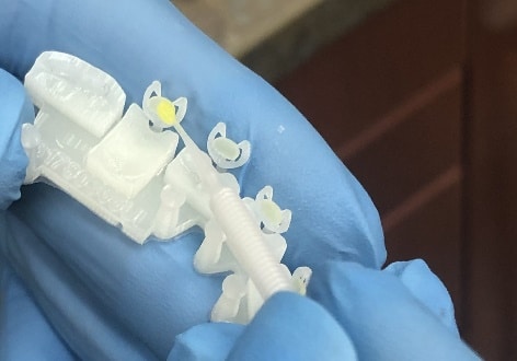
As with most dental bonding procedures, one of the keys to efficient and successful bonding of Specifix attachments is dry field isolation. Additionally, it is important to avoid adhesive contamination of the attachment positioners, the breakaway features, and the frames that connect the attachments to the jig. The adhesive should be brushed onto the attachment base immediately before seating the positioning jig, and not ahead of time (ie, do not place adhesive on the Specifix attachments before the patient arrives). We use a 1-mm superfine micro brush to ensure accurate and consistent placement of the bonding agent. We also do not light cure the brushed adhesive before seating the attachments, because we want the adhesive layer to be as thin as possible for maximum dimensional accuracy of the bonded attachment.
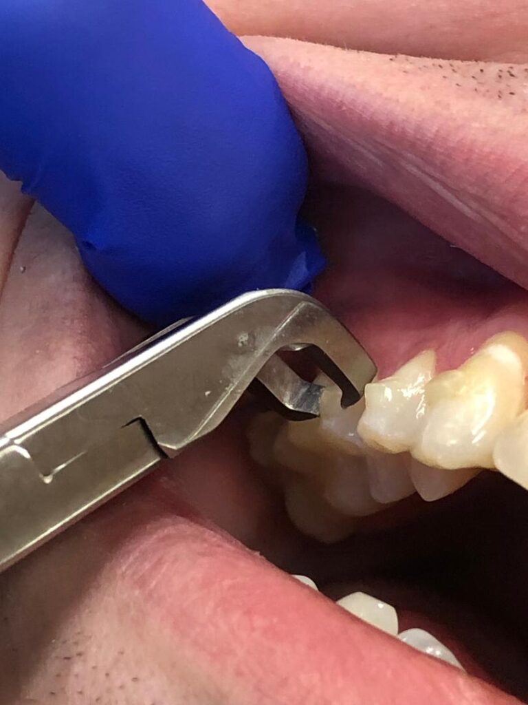
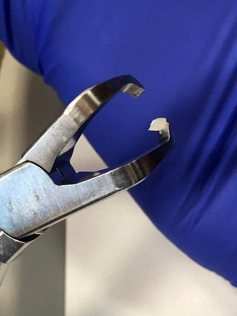
Removal of Specifix attachments is a quick and easy procedure. We prefer removing Specifix attachments using angled bracketremoval pliers which are designed to safely debond orthodontic brackets in one piece. In our experience, this instrument performs quite well for removing Specifix attachments.
Summary
We have observed that Invisalign Specifix attachments are designed to deliver durable, dimensionally stable attachment surfaces that optimize aligner engagement from start to finish. The material blends well with the patient’s natural tooth color, preserving excellent esthetics throughout Invisalign treatment.
This innovation of 3D printed attachments to the precise shape, size, and accurate location per the doctor’s ClinCheck treatment plan results in a streamlined bonding workflow with no composite flash clean up.
At the completion of treatment, the attachments can be removed cleanly and efficiently with angled bracket removal pliers. OP
Photos courtesy of Dr Manal Ibrahim LaVacca.
Footnote:
- Manufacturer’s note: Lingual Specifix attachments are not available at the time of this publication, but an attachment template will be provided if these are part of the patient’s treatment plan.

Manal Ibrahim LaVacca, DDS, is a Diplomate of the American Board of Orthodontics and a dual‑trained specialist in prosthodontics and orthodontics. She founded Innovative Orthodontic Centers in Illinois and, with her husband, prosthodontist Anthony LaVacca, DMD, MS, FACP, FICOI, co‑founded Innovative Dental Partners, a multispecialty group providing prosthodontic, pediatric, general, and specialty care. She earned her dental and prosthodontic degrees from the University of Illinois and completed her orthodontic training at New York University. Ibrahim can be reached at: [email protected]