 Christopher W. Scharff is the vice president of sales at Imaging Sciences International Inc in Hatfield, Pa. He has been with the company for more than 8 years. As vice president of sales, Scharff guides and supports the entire Imaging Sciences sales team by proposing and executing policies and programs to achieve maximum sales-volume potential. Scharff also oversees all of the marketing efforts in the promotion of the i-CAT and coordinates the schedule for all industry trade shows and meetings.
OP: What types of tomographic imaging systems does your company provide to the dental community?
Scharff: Imaging Sciences International develops and manufactures computer-controlled dental and maxillofacial radiography products, including the CommCAT, the Panorex CMT, and the i-CAT. These on-site imaging units provide predictable treatment outcomes to dentists and other health care professionals.
OP: Which of your systems is the most popular? Why?
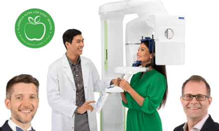
Scharff: Our most popular system is the Imaging Sciences i-CAT. The easy-to-use i-CAT is the leader in cone-beam 3-D imaging and produces thorough 3-D views of oral and maxillofacial structures. Patients sit in an “open environment scan,” which increases patient comfort and captures the natural orientation of anatomy. The data is transferred to a computer, typically within 2 minutes, and is displayed on a 3-D mapping tool that allows doctors and technicians to easily format and select desired slices for immediate viewing. The i-CAT workstation, including the machine, computer station, and access space, requires less than 60 square feet of office space, and creates 3-D images with less radiation than traditional fan-beam computed tomography systems.
OP: How does the i-CAT benefit orthodontists?
Scharff: With an extended field of view option, the i-CAT, which captures 22 cm of height (nearly twice the amount of information that the 13-cm traditional scans capture), orthodontists create a complete orthodontic workup of frontal and lateral cephalometric, panoramic, supernumerary, SMV, TM joint, and airway and spinal studies. The i-CAT scan, a 14-bit system that produces images with 16,384 shades of gray, allows specialists to capture the images needed for cephalometric reconstruction and to provide accurate treatments and predictable outcomes of orthodontic procedures and oral surgeries. The system is compatible with Dolphin 3D and 3dMD software and provides 3-D volume-rendering software for viewing and rotating data from all angles.


