by Neal D. Kravitz, DMD, MS
A noncompliant alternative to Phase I mandibular expansion
 |
| Neal D. Kravitz, DMD, MS |
Crowding is the most prevalent component of malocclusion among dental patients. Early interceptive (or Phase I) orthodontic expansion in the mixed dentition can prevent unnecessary loss of arch length, restore occlusal function, and possibly minimize the need for premolar extractions during adolescence. This article will review the proper use of the mandibular E-arch (expansion arch, Arnold appliance) during Phase I treatment.
Crowding: Prevalence, Etiology, and Signs
Crowding, otherwise referred to as a tooth-size/arch-length discrepancy (TSALD), is the consequence of a disharmony between tooth size and the space available in the dental arch.1 According to the National Center for Health Statistics (Washington, DC), 40% of children (aged 6 to 11)2 and 85% of adolescents (aged 12 to 17)3 have crowding problems.
The etiology of TSALD is multifactorial due to both hereditary and environmental causes.1 Hereditary factors include large tooth size, small arch length, narrow arch width, supernumerary or missing teeth, and abnormal crown morphology.4 These factors may be attributed to the evolutionary development of modern humans and a progressive reduction in jaw size. Environmental influences include disturbances in dental eruption, premature loss of deciduous teeth, interproximal caries in deciduous teeth, transposition, muscle imbalance, and socioeconomic conditions.4
 |
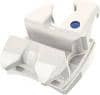
| Figure 1: A mandibular E-arch. (AOA Orthodontic Appliances). Compression of the Elgiloy coil spring during seating of the appliance allows for posterior dentoalveolar decompensation and distalization of the first molars. If anterior brackets are to be placed, ask the lab to keep the buccal tubes on the molar bands. |
An early sign of a developing TSALD is the absence of spacing in the primary dentition. A “closed”5 primary dentition prohibits the early mesial shift (the mesial migration of the erupting permanent mandibular first molar into a Class I relationship, closing the lower primate space) and fails to account for incisor liability (the size differential between the primary and permanent incisors). In the early mixed dentition, clinical signs of crowding may include dentoalveolar protrusion without interproximal spacing, overlapping incisors, or premature exfoliation of the deciduous canines.4
Debate Over Mandibular Expansion
According to McNamara,6 TSALD can be simplified into three categories: clear-cut extraction patients (mandibular crowding >6 mm), clear-cut nonextraction (mandibular crowding <3 mm), and borderline crowding patients (mandibular crowding 3 to 6 mm). For borderline patients with moderate crowding, deciding whether to regain space with mandibular expansion or to begin serial extraction can be challenging.
Orthopedic expansion in the mandibular arch is limited because of the lack of a midline suture. Mandibular expansion primarily induces tooth inclination localized to the dentoalveolus, and these effects are thought to relapse. As such, mandibular intercanine dimension is purported to be an inviolable measurement.
Despite its clinical importance, few studies have examined the effects of mandibular expansion. Motoshi et al7 reported a 5.42-mm (standard deviation 1.98) increase in intermolar width and 10.16º (standard deviation 3.83) of buccal tooth inclination during early expansion with the Schwarz appliance. Housley et al8 reported that transverse expansion was more stable in the posterior region of the mandibular dental arch than in the anterior region. Prospective clinical data9 from the Michigan Expansion Study (MES) has shown long-term stability of mandibular expansion in borderline crowding patients when mandibular expansion is combined with maxillary rapid palatal expansion during the early mixed dentition. However, questions still remain as to whether violating intercanine dimension relegates the patient to a lifetime of retention.
 |
| Figure 2: (Left) Two orthodontic elastics are used to compress the NiTi coil spring. (Center) Bending the E-arch “downward” may help seat the frame below the occlusal surface of the posterior teeth. (Right) A properly seated E-arch. The elastics are cut away with a pin and ligature cutter. |
The E-arch
The E-arch is a fixed, spring-loaded expander for slow expansion of the maxillary or mandibular arches. Developed by Arnold, and popularized by Berkowitz, the E-arch was invented for maxillary orthopedic expansion in patients with cleft palates. In the mandible, the E-arch can create a modest amount (4 to 5 mm) of arch space in the lower anterior region by orthodontic tipping (rather than orthopedic change) of the lower posterior teeth, as well as distalization of the first molars.
The appliance consists of a 0.040-inch tube-like frame with a 0.010- x 0.040-inch Elgiloy or NiTi open coil spring (Figure 1). Seating the E-arch compresses the open coil spring and activates expansion. Therefore, the E-arch is ideal where patient compliance or parent cooperation is a concern. The E-arch is typically banded to the first permanent molars; however, if these teeth have not fully erupted, bands can be placed on the second deciduous molars. In these situations, the clinician may consider placing lingual extension arms to prevent the second deciduous molars from expanding further than the first permanent molars.
 |
| Figure 3: Bonding the tube-frame to the teeth. After 2 months, the patient began lifting the lingual frame up with her tongue, causing the frame to rest on top of the first primary molars. The frame was bent “downward” intraorally using three-prong pliers. RM Bond flowable composite was placed on the primary left canine to secure the tube-frame. Note the significant amount of molar distalization. |
If you think that incisor brackets are needed, inform the laboratory to keep the buccal tubes on the molar bands. Severe incisor malrotation, deep overbite, anterior crossbite, or early loss of the mandibular deciduous canines may warrant incorporation of a 2 x 4 for incisor alignment or advancement. In particular, early loss of the mandibular deciduous canines results in lingual migration of the permanent incisors, blocking eruption of the mandibular canines and preparing the patient for serial extraction.
Getting Started
First, wrap one or two orthodontic elastics around the frame of the E-arch to help compress the coil spring. Remove the patient’s spacers and seat the appliance without cement to test for proper fit. The frame should seat below the occlusal surface of the deciduous molars. The clinician may consider bending the frame slightly “downward” to prevent the appliance from climbing over the teeth during expansion. After the bands are luted (I use GC Fuji Ortho Band LC Automix) and the appliance is fully seated, remove the orthodontic elastics with a pin and ligature cutter (Figure 2).
The mandibular E-arch can be left active for approximately 6 months, depending on the amount of expansion needed. For patients requiring expansion in both arches, dentoalveolar decompensation with the mandibular E-arch establishes a “reference” mandibular arch width to guide the amount of maxillary expansion.9 Some patients have a tendency to try to flick the lingual frame with their tongue or pull on the appliance with their fingers. In these situations, consider bonding the lingual frame (not the coil spring) to a posterior tooth for added stability (Figure 3). While bonding the lingual frame will not affect symmetrical expansion, it may impeded equal bilateral molar distalization.
Once you have achieved adequate mandibular expansion, you have the option of either removing the E-arch and delivering a lower Hawley or deactivating the coil spring and converting the appliance into a holding arch. The E-arch is deactivated by sectioning the coil spring intraorally using a FG #557 straight fissure crosscut carbide bur. For patient comfort, you may consider placing a small amount of flowable composite (such as RM Bond from Rocky Mountain Orthodontics) over the sectioned coil spring. The “passive” E-arch can be left on until the start of Phase II or the eruption of the second premolars (Figure 4).
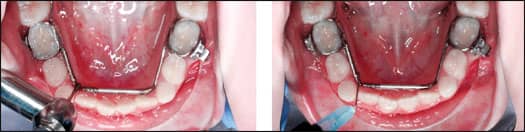 |
| Figure 4: Deactivation of the mandibular E-arch. The patient presented with fully blocked-out lateral incisors. (Left) Expansion was discontinued after 6 months by sectioning the coil spring with a FG #557 straight fissure crosscut carbide bur. (Right) Flowable composite was placed over the coil spring for comfort. Note the lingual extension arms to aid expansion of the first permanent molars. |
 |
| Figure 5: A blocked-out lower right lateral incisor. (Left) Delivery of a Schwarz appliance. Patient was noncompliant with appliance wear and required numerous appliance-adjustment appointments. (Right) The same patient after 8 months of E-arch wear. After adequate expansion was achieved, the coil spring was deactivated and flowable composite was placed for comfort. Note the excessive buccal tipping of the first molars, a consequence of prolonged use of the E-arch. |
The E-arch offers numerous advantages in comparison to other popular methods of Phase I mandibular expansion, such as the Schwarz appliance and the lip bumper:
- It offers noncompliant expansion and molar distalization;
- during treatment, no appliance adjustment appointments are needed;
- it allows for the easy incorporation of incisor brackets; and
- after expansion, the appliance easily converts into a holding arch, providing noncompliant retention until Phase II (Figure 5).
The primary disadvantage of the mandibular E-arch is excessive buccal tipping of the posterior teeth if left unmonitored. Lower dentoalveolar expansion may also result in dental extrusion and increased lower anterior facial height.10 Additionally, in patients with poor oral hygiene, the frame can accumulate calculus or become imbedded in the soft tissue. If this occurs, you should section the lingual frame and complete Phase I treatment with a 2 x 4.
Conclusion
The mandibular E-arch offers noncompliant dentoalveolar decompensation and molar distalization for preadolescent patients with moderate crowding. By intervening during the mixed dentition, orthodontists can eliminate potential irregularities and aid dental eruption. The decision whether to expand or extract, however, requires proper diagnosis and must be made on a case-by-case basis.
Neal D. Kravitz, DMD, MS, is in private practice in South Riding, Va, and White Plains, Md. He is a diplomate of the American Board of Orthodontics, and is on the faculty at the University of Maryland and Washington Hospital Center. He can be reached at or kravitzorthodontics.com.
References
- Ngan P, Alkire RG, Fields H Jr. Management of space problems in the primary and mixed dentitions. J Am Dent Assoc. 1999;130:1330-1339.
- Kelly JE, Sauchey M, VanKirk LE. An assessment of the occlusion of teeth of children. Washington, DC: National Center for Health Statistics; 1973. Department of Health Education and Welfare publication (HRA) 74-1612.
- Kelly JE, Harvey C. An assessment of the occlusion of teeth of youths 12-17 years. Washington, DC: National Center for Health Statistics; 1973. Department of Health Education and Welfare publication (HRA) 77-1644.
- Dale JG. Guidance of occlusion: serial extraction. In: Graber TM, Swain BF, eds. Orthodontics: Current principles and techniques. St Louis: Mosby;1985:259-366.
- Baume LJ. Physiological tooth migration and its significance for the development of occlusion. I. The biogenetic course of the deciduous teeth. J Dent Res. 1950;29(4):440-447.
- McNamara JA Jr. Early intervention in the transverse dimension: is it worth the effort? Am J Orthod Dentofacial Orthop. 2002;121(6):572-574.
- Motoyoshi M, Shirai S, Yano S, Nakanishi K, Shimizu N. Permissible limit for mandibular expansion. Eur J Orthod. 2005;27(2):115-20.
- Housley JA, Nanda RS, Currier GF, McCune DE. Stability of transverse expansion in the mandibular arch. Am J Orthod Dentofacial Orthop. 2003;124(3):288-293.
- O’Grady PW, McNamara JA Jr, Baccetti T, Franchi L. A long-term evaluation of the mandibular Schwarz appliance and the acrylic splint expander in the early mixed dentition patients. Am J Orthod Dentofacial Orthop. 2006;130(2):202-213.
- Wendling LK, McNamara JA Jr, Franchi L, Baccetti T. A prospective study of the short-term treatment effects of the acrylic-splint rapid maxillary expander combined with the lower Schwarz appliance. Angle Orthod. 2005;75(1):7-14.