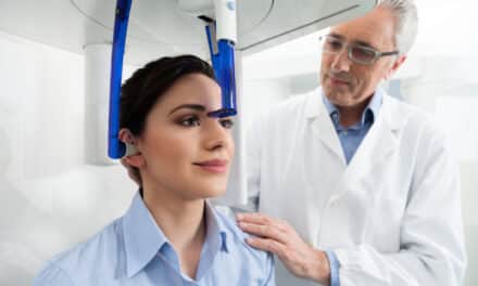
The first step in diagnosis is a clear picture
Planmeca Inc
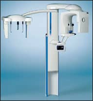
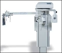
Planmeca Inc, Roselle, Ill, offers the ProMax 3D x-ray with Cone Beam Volumetric Tomography (CBVT), which features 3D and digital panoramic imaging; digital panoramic bitewings; and digital cephalometrics, all in one system. ProMax 3D has a small footprint for on-site use in any office. The ProMax 3D x-ray has an effective exposure time of 6 seconds, with a scan rate of 16 to 18 seconds per image. Planmeca also offers a software solution for 3D users. Romexis acquisition software allows manual artifact removal only when necessary, maintaining the integrity of the original volume. This data can then be transferred to N-Liten or any DICOM-compatible 3D rendering software. N-Liten 3D is available with the new N-Liten Implant Module, which aids in the placement of implants.
Planmeca Inc
(630) 529-2300
www.planmecausa.com

PracticeWorks
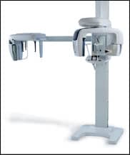
PracticeWorks, Atlanta, introduces the KODAK 9000C 3D Extraoral Imaging System, which allows orthodontists to obtain low-dose, high-resolution, 3D images, as well as panoramic and cephalometric images.Panoramic image quality is optimized for the jaw morphology with the new variable focal trough feature. The three-in-one system captures a localized field of view 3D image, which can be limited to, for example, an impacted cuspid area, the roots of the upper centrals, or a temporomandibular joint area. The KODAK 9000C system requires 20% less space than its predecessor, the KODAK 8000C panoramic and cephalometric system. Its motorized collimator offers a broad range of cephalometric formats. It suits any orthodontic tracing need, from the full skull (12- x 12-inch) to standard (8- x 10-inch), and a small field for lower dose exposures. The system also generates lateral, frontal, and submental-vertex images.
PracticeWorks
(800) 944-6365
www.kodakdental.com/go/ortho

Imaging Sciences International
Imaging Sciences International, Hatfield, Pa, offers the i-CAT® Cone Beam 3D dental imaging system, which has been redesigned with enhanced features for patient diagnosis, treatment, and surgical predictability. The larger field of view captures anatomically accurate and detailed cephalometric 3D images of the entire skull. Views range from 4 cm to 17 cm in height and 16 cm to 23 cm in diameter. The amorphous silicon flat-panel sensor adjusts to capture data in portrait and landscape view. The i-CAT scans take only 5, 8.9, and 26 seconds, with reconstruction of a typical scan in less than 30 seconds. File sizes are less than 50MB.
Imaging Sciences International
(800) 205-3570
www.i-cat.com

Sirona Dental Systems
Sirona Dental Systems, Charlotte, NC, introduces the Galileos 3D Cone Beam Imaging system. Galileos combines x-ray diagnostics, 3D visualization, treatment planning, and patient communication in one unit. Single scan capability captures a comprehensive scan of a patient’s oral/maxillofacial region at the lowest dose of radiation. The familiar panoramic view has a unique, real-time evaluation window to scroll through 3D anatomy. The integrated diagnosis and planning system provides workflow integration from a quick scan to treatment planning. Galileos may be integrated with current third-party software programs.
Sirona Dental Systems LLC
(800) 659-5977
www.sirona.com

Gendex Dental Systems
Gendex Dental Systems, Des Plaines, Ill, introduces the Orthoralix® 9200 DDE, which combines the imaging technology of the Orthoralix 9200 AEC+ with the latest-generation sensor. At the core of the system is the CCD sensor module, which replaces the traditional film cassette, eliminating the hassles and cost of a processor, film, and chemicals.
The 9200 DDE includes basic and advanced imaging programs and automatic exposure control, designed to produce high-quality images. Available upgrades include cephalometric imaging and TRANSCAN cross-sectional tomography.
Gendex Dental Systems
(888) 275-5286
www.gendex.com

Air Techniques
Air Techniques Inc, Melville, NY, offers the ScanX® Digital Imaging system, a simple solution for upgrading to digital. The system uses existing x-ray equipment to create digital images that are ready within seconds. View images in as little as 4 seconds or an entire full mouth series in less than 2 minutes. Scan and automatically erase Phosphor Storage Plates (PSP) in the same continuous cycle using In-Line Erase. ScanX features Intelligent Track Control, which allows two users, treating different patients to operate ScanX simultaneously. The PSPs are available in sizes zero through four, in panoramic and cephalometric, and are reusable thousands of times. ScanX is available in three different models: Classic, for intraoral and extraoral imaging; ScanX Intraoral, for central image processing; and compact ScanX Duo, for chairside intraoral imaging.
Air Techniques Inc
(800) AIR-TECH
www.airtechniques.com
J. Morita
J. Morita, Irvine, Calif, introduces the Veraviewepocs 2D/3D, which offers digital 3D, panoramic, and cephalometric imaging options. The 2D model can be upgraded to 3D at any time. The multifunction cassette allows operators to take panoramic and 3D images without changing the cassette. For 3D positioning, simply click the region of interest on the panoramic view and the machine automatically readjusts for the perfect shot.
J. Morita
(877) JMORITA
www.jmoritausa.com
Panoramic Corp
Panoramic Corp, Fort Wayne, Ind, offers panoramic and cephalometric x-ray systems. With panoramic, TMJ, and cephalometric imaging, the free-standing PC-1000/Laser 1000 provides radiographic capability at an affordable price with the industry’s best warranty, according to the company. Combine it with a daylight load processor, and you have a darkroom without the darkroom.
Panoramic Corp
(800) 654-2027
/b>

AFP Imaging
AFP Imaging Corp, Elmsford, NY, introduces the latest version of its NNT software. NNT is used in conjunction with the NewTom family of cone beam 3D imaging products for advanced 3D dentomaxillofacial imaging. NNT 2.13 offers user-friendly options to help in collaborating with other professionals. NNT 2.13 allows users to import DICOM data sets that were generated using a NewTom 3D scanner. NNT 2.13 exports DICOM images as a single file, reducing file space, and comes with free viewing capabilities. The free viewer, now standard with version 2.13 and higher, allows practitioners to view images exported with DICOM without purchasing a full version of the NNT software. Image data can be burned directly onto a CD or DVD. The image-sharing capabilities, coupled with reporting available in a pdf format, improve the workflow among imaging specialists, dental laboratories, and clinicians. NNT 2.13 allows direct integration with third-party software programs.
AFP Imaging Corp
(800) 592-6666
www.afpimaging.com

Dexis
Dexis LLC, Des Plaines, Ill, offers the Dexis® Digital X-ray System, which features “One-Click FMS,” a ClearVu™ image enhancement tool, and a PerfectSize™ sensor. The software allows clinicians to customize “capture” and “display” features as well as reports and letters. Expanded communication options permit images to be shared in a variety of formats. Dexis also provides integration with practice-management programs, digital pan systems, cameras, and scanners to create an “Imaging Hub” that allows the orthodontist to capture, store, and access intraoral and extraoral radiographic and camera images for maximum productivity.
Dexis
(888) 883-3947
www.dexis.com