A New View
Michelle Jackson, Imaging Sciences International
 |
Michelle Jackson is marketing manager at Imaging Sciences International, Hatfield, Pa. She oversees all of the marketing and branding initiatives for The Next Generation i-CAT®, Imaging Sciences’ flagship product.
OP: Please describe the features of the Next Generation i-CAT.
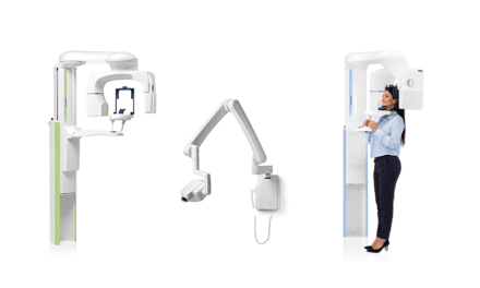
Jackson: It is a cone beam 3D dental imaging technology. The system has a sleek new design and enhanced features for treatment, diagnosis, and surgical predictability. The i-CAT offers an extended field of view and captures anatomically accurate and detailed cephalometric 3D images of the entire skull—up to 17 cm in height and 23 cm in diameter. An amorphous silicon flat-panel sensor can be adjusted to capture data in two views—portrait and landscape—with the ability to collimate for a range of volumes. The standard i-CAT scan takes only 8.9 seconds, and reconstruction takes less than 30 seconds. File sizes are less than 50MB, allowing them to be manageable for file and case sharing.
OP: How does your i-CAT Cone Beam 3D Imaging System enhance the quality of care and increase predictability of treatment outcomes for orthodontic procedures?
 |
Jackson: The i-CAT improves the accuracy of orthodontic diagnosis, treatment planning, monitoring treatment response, and treatment outcome evaluation. The scan exposes pathologies and other issues that are undetectable with traditional methods, giving orthodontic professionals a better insight into the relationship between patients’ teeth, jaw, nerves, bone, and underlying dental structures—which can lead to discovering the unexpected well in advance of a procedure.
The extended field of view option was specifically designed for orthodontic imaging and is ideal for cephalometric reconstruction. In just one i-CAT scan, you can create a complete orthodontic workup, including cephalometrics, SMV, supernumerary, airway and spinal views, panoramic, TMJs, and impactions. The i-CAT allows you to easily locate impacted canines and supernumeraries and check for root resorption. It provides distortion-free 3D views of critical anatomy surrounding the condyles and the tools to accurately measure and determine the proper course of treatment.
OP: Does your company provide continuing education classes?
Jackson: All i-CAT customers are provided with a 11/2-day hands-on, in-office training, which is included with the purchase of an i-CAT. The training includes instruction on how to take a scan, proper patient positioning, postprocessing of data (navigating and mapping anatomy), exporting data, creating reports, and information sharing.
Imaging Sciences also offers training on the proprietary i-CAT Vision software that comes equipped with every i-CAT imaging system. i-CAT Vision is free software that optimizes practice workflow with tools for unlimited networking and sharing of patient scans.
We also launched the i-CAT Institute™ Education and Training Center, which is available to all i-CAT customers as well as any other orthodontist who is interested in learning about Cone Beam 3D technology. The new state-of-the-art institute provides instruction on the use of Cone Beam 3D volume data and data manipulation, DICOM-compliant third-party software, and other topics relating to the industry’s growing demands. More information on the courses offered at the training center can be found at www.i-CAT3D.com.
OP: Is the i-CAT compatible with third-party orthodontic planning software?
Jackson: The i-CAT is DICOM-3-compatible, so it is able to seamlessly integrate with a number of third-party applications, such as Dolphin®, 3dMd, InVivo, and CyberMed. A full list can be found at www.i-CAT.com.
|
For more information about Imaging Sciences, see our online Buyer’s Guide. |
The i-CAT recently became the first cone beam 3D imaging system to receive certification for compatibility with SureSmile Technology, which increases treatment accuracy and precision and reduces treatment time. Orthodontists can take an i-CAT scan of the patient’s mouth, face, and jaw, and use the data in the SureSmile system to control the treatment through virtual diagnostic simulations, instant quality grading tools, prescriptive planning capabilities, and robotic archwire customization.